. 2025 Jan 7;14(2):62.
doi: 10.3390/cells14020062.

The MCPH7 Gene Product STIL Is Essential for Dendritic Spine Formation

Affiliations

The MCPH7 Gene Product STIL Is Essential for Dendritic Spine Formation

Tohru Matsuki et al. Cells. .

Abstract

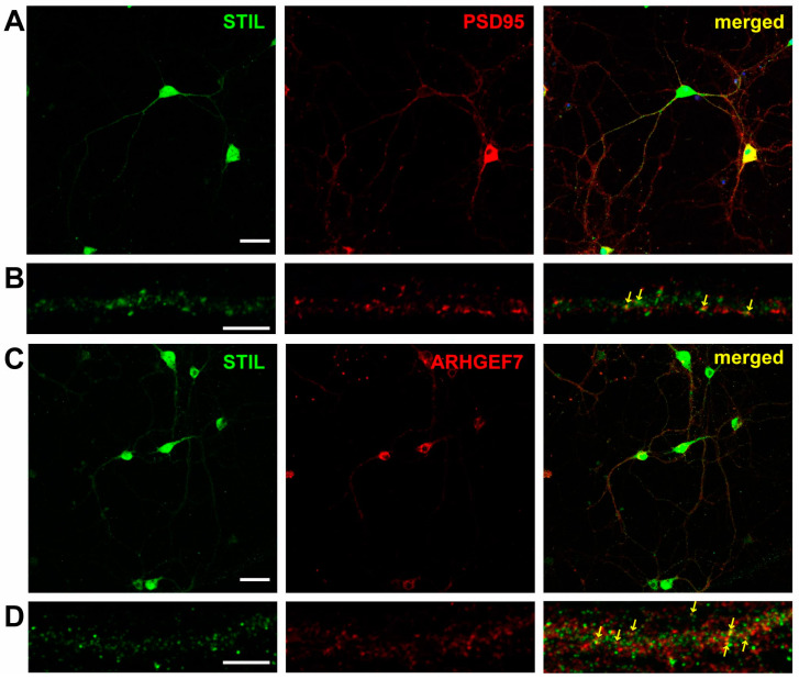
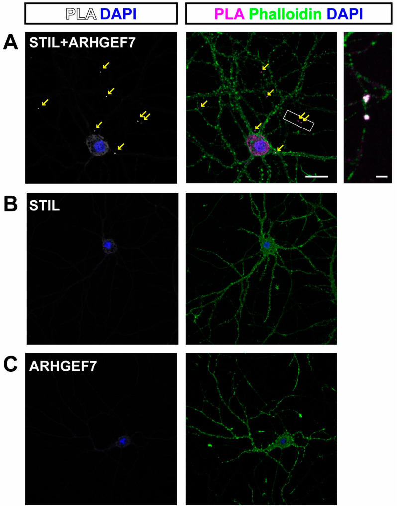
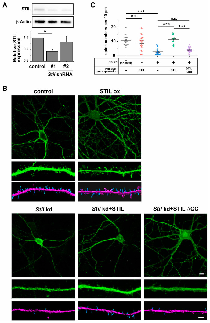
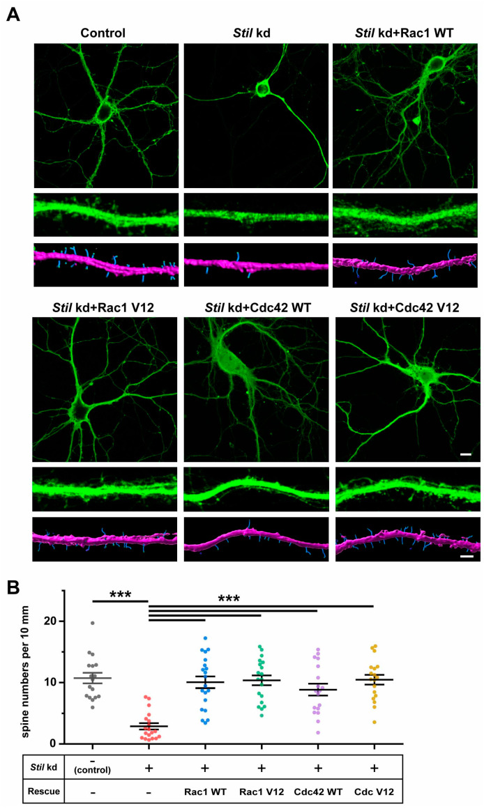
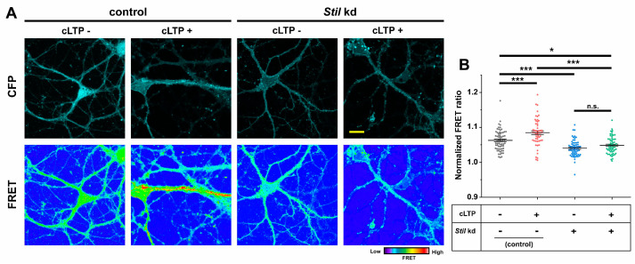
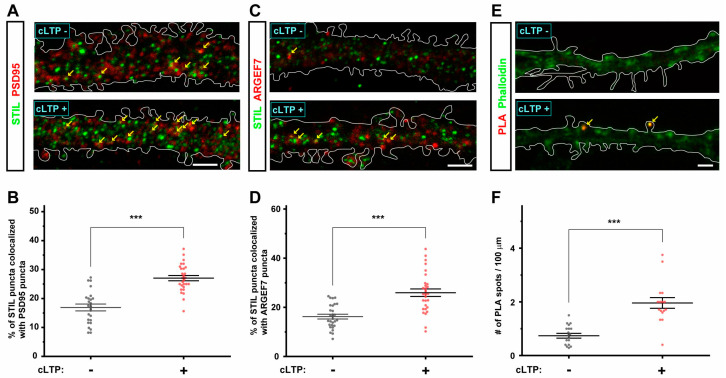
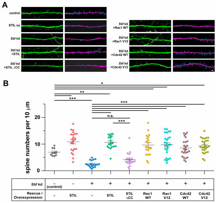
Dendritic spine formation/maintenance is highly dependent on actin cytoskeletal dynamics, which is regulated by small GTPases Rac1 and Cdc42 through their downstream p21-activated kinase/LIM-kinase-I/cofilin pathway. ARHGEF7, also known as ß-PIX, is a guanine nucleotide exchange factor for Rac1 and Cdc42, thereby activating Rac1/Cdc42 and the downstream pathway, leading to the upregulation of spine formation/maintenance. We found that STIL, one of the primary microcephaly gene products, is associated with ARHGEF7 in dendritic spines and that knockdown of Stil resulted in a significant reduction in dendritic spines in neurons both in vitro and in vivo. Rescue experiments indicated that the STIL requirement for spine formation/maintenance depended on its coiled coil domain that mediates the association with ARHGEF7. The overexpression of Rac1/Cdc42 compensated for the spine reduction caused by STIL knockdown. FRET experiments showed that Rac activation is impaired in STIL knockdown neurons. Chemical long-term potentiation, which triggers Rac activation, promoted STIL accumulation in the spine and its association with ARHGEF7. The dynamics of these proteins further supported their coordinated involvement in spine formation/maintenance. Based on these findings, we concluded that the centrosomal protein STIL is a novel regulatory factor essential for spine formation/maintenance by activating Rac and its downstream pathway, possibly through the association with ARHGEF7.

Keywords: ARHGEF7; Cdc42; MCPH7; Rac1; STIL; dendritic spine.


Conflict of interest statement

The authors declare that the research was conducted in the absence of any commercial or financial relationships that could be construed as a potential conflict of interest.

Figures

Figure 1
Figure 1
STIL co-localizes with PSD95 and ARHGEF7 along dendrites of cultured hippocampal neurons. (A) Representative images of hippocampal neurons stained for STIL (green) and PSD95 (red). (B) High magnification views of STIL and PSD95 distribution in a dendrite. Co-localization of STIL and PSD95 signals is indicated by arrows in the merged image. (C) Representative images of hippocampal neurons stained for STIL (green) and ARHGEF7 (red), along with a merged image. (D) High magnification views of STIL and ARHGEF7 distribution in a dendrite. Co-localization of STIL and ARHGEF7 signals is indicated by arrows in the merged image. Scale bars = 50 µm for (A,C) and scale bars = 5 µm for (B,D).
Figure 2
Figure 2
STIL associates with ARHGEF7 in dendritic spines of cultured hippocampal neurons. (A) Representative images of cultured hippocampal neurons subjected to proximity ligation assay (PLA). PLA signals originating from the proximity of STIL and ARHGEF7 are detected as red spots and are indicated by arrows (left). A green phalloidin-stained image is overlaid on the PLA image to show dendritic spines (middle). The PLA signals appear as white or magenta spots, indicating that they localize to the dendritic spine. The right image shows a magnified view of the boxed area in the middle. Scale bars = 20 µm (middle image) and 2 µm (right image). (B,C) Representative images of cultured hippocampal neurons in negative control experiments. No PLA signal is observed when antibodies against STIL (B) and ARHGEF7 (C) are applied independently.
Figure 3
Figure 3
Stil knockdown causes dendritic spine reduction in cultured hippocampal neurons, which is rescued by knockdown-resistant human STIL but not by human STIL lacking the coiled coil domain. (A) Confirmation of Stil knockdown by Stil shRNAs. Proteins were extracted from cultured neurons infected with lentiviral vectors expressing Stil shRNA or control empty vectors, and STIL expression levels were examined by Western blotting in four independent experiments. A representative STIL band image is shown at the top, and an anti-β-Actin band image as loading control is shown at the middle. The signal intensity of the STIL band was corrected against the signal intensity of the β-Actin band, and a graph of the values compared to the control is shown at the bottom. * p < 0.01 (B) Representative images of a neuron overexpressing STIL and Stil knockdown neurons introduced with/without rescue vectors. Raw images of whole cells (top panel) and magnified dendrites (middle panel) and reconstructed images of dendrites (bottom panel) of hippocampal neurons cultured under various experimental conditions are shown. EGFP alone (control), STIL (STIL ox), Stil shRNA #1 (Stil kd), Stil shRNA #1 with knockdown-resistant STIL (Stil kd + STIL), or Stil shRNA #1 with knockdown-resistant STIL lacking a coiled coil domain (Stil kd + STIL ∆CC) was introduced into neurons. The spines and dendritic shafts are visualized in blue and pink, respectively, in the reconstructed images. Scale bars = 10 µm (top panel) and 2 µm (bottom panel). (C) Distribution of dendritic spine density (number of spines per 10 µm of dendrite) under each condition. Each dataset was obtained from dendrites in neurons analyzed in at least three experiments. Mean values: 10.721 (control, n = 17), 9.637 (STIL WT, n = 22), 8.158 (STIL ∆CC, n = 22), 2.877 (Stil kd, n = 19), 11.188 (Stil kd + STIL WT, n = 13), and 4.005 (Stil kd + STIL ∆CC, n = 20). *** p < 0.001. n.s., not significant.
Figure 4
Figure 4
Rac1 and Cdc42 induction can compensate for the reduction in dendritic spines caused by Stil knockdown. (A) Representative images of control and Stil knockdown neurons with/without expressing wild-type or constitutively active Rac1/Cdc42. Raw images of whole cells (top panel) and of magnified dendrites (middle panel) and reconstructed images of dendrites (bottom panel) of cultured hippocampal neurons under various experimental conditions are shown. Stil shRNA #1 with wild-type Rac1 (Stil kd + Rac1 WT), Stil shRNA #1 with constitutively active Rac1 (Stil kd + Rac1 V12), Stil shRNA #1 with wild-type Cdc42 (Stil kd + Cdc42 WT), or Stil shRNA #1 with constitutively active Cdc42 (Stil kd + Cdc42 V12) was introduced into neurons. The spines and dendritic shafts are visualized in blue and pink, respectively, in the reconstructed images. Scale bars = 10 µm (top panel) and 2 µm (bottom panel). (B) Distribution of dendritic spine density (number of spines per 10 µm of dendrite) under each condition. The data for the control and Stil kd alone are the same as those shown in Figure 3C. Each dataset was obtained from dendrites in neurons analyzed in at least three experiments. Mean values: 10.721 (control, n = 17), 2.877 (Stil kd, n = 19), 10.048 (Stil kd + Rac1 WT, n = 20), 10.357 (Stil kd + Rac1 V12, n = 26), 8.853 (Stil kd + Cdc42 WT, n = 25), and 10.454 (Stil kd + Cdc42 V12, n = 26). *** p < 0.001 Data were obtained from three independent experiments.
Figure 5
Figure 5
Stil knockdown impairs Rac activation in cultured hippocampal neurons. Raichu-Rac1, a bioindicator of Rac activity, was introduced into control and Stil kd neurons, FRET ratios were visualized, and FRET efficiencies were calculated. (A) Representative images showing CFP signal (upper panels) and FRET ratio (YFP/CFP; lower panels) in cultured hippocampal neurons harboring Raichu-Rac1. Control neurons with and without cLTP induction (cLTP+ and cLTP−, respectively), along with Stil kd neurons with and without cLTP induction, are shown. Scale bar = 5 µm. (B) Distribution of normalized FRET ratio under each condition. Each dataset was obtained from dendrites in neurons analyzed in at least three experiments. Mean values: 1.063 (control, cLTP−, n = 67), 1.085 (control, cLTP+, n = 53), 1.040 (Stil kd, cLTP−, n = 63), and 1.049 (Stil kd, cLTP+, n = 59). * p < 0.05, *** p < 0.001, and n.s.: not significant.
Figure 6
Figure 6
cLTP induces the accumulation of STIL in spines and promotes the association of STIL with ARHGEF7. (A) Representative images showing the distribution of STIL (green) and PSD95 (red) in the dendrite of cultured hippocampal neurons with and without cLTP induction (cLTP+ and cLTP−, respectively). The dendrites are outlined with white lines. Co-localization of STIL and PSD95 signals is indicated by arrows. Scale bar = 5 µm. (B) Ratios of STIL puncta co-localizing with PSD95 puncta among all STIL puncta in the dendrite of cultured hippocampal neurons with and without cLTP induction. Mean values: 16.902 (cLTP−, n = 24) and 27.042 (cLTP+, n = 28). (C) Representative images showing the distribution of STIL (green) and ARHGEF7 (red) in the dendrite of cultured hippocampal neurons with and without cLTP induction. The dendrites are outlined with white lines. Co-localization of STIL and ARHGEF7 signals is indicated by arrows. Scale bar = 5 µm. (D) Ratios of STIL puncta co-localizing with ARHGEF7 puncta in the dendrite of cultured hippocampal neurons with and without cLTP induction. Mean values: 16.219 (cLTP−, n = 27) and 25.943 (cLTP+, n = 29). (E) Representative images showing PLA signal spots (red) in the dendrite of cultured hippocampal neurons with and without cLTP induction. Spines are stained with phalloidin (green). The dendrites are outlined with white lines. PLA signals in the spine are indicated by arrows. Scale bar = 2 µm. (F) PLA signal spot density (numbers of PLA signal spots/100 µm) in the dendrite of cultured hippocampal neurons with and without cLTP induction. Mean values: 0.737 (cLTP−, n = 19) and 1.959 (cLTP+, n = 16). Each dataset shown in (B,D,F) was obtained from dendrites in neurons analyzed in at least three experiments. *** p < 0.001.
Figure 7
Figure 7
In vivo Stil knockdown causes dendritic spine reduction in cerebral cortical neurons, which is rescued by knockdown-resistant human STIL but not by human STIL lacking the coiled coil domain. Rac1 and Cdc42 induction can compensate for the reduction in dendritic spines caused by in vivo Stil knockdown. (A) Representative images of dendrites of a neuron overexpressing STIL and Stil knockdown neurons introduced with/without various rescue vectors. Each image set shows a raw image (left) and a reconstructed image (right) of a dendrite of a mouse cortical neuron at P30. EGFP alone (control), STIL (STIL ox), Stil shRNA (Stil kd), Stil shRNA with knockdown-resistant STIL (Stil kd + STIL WT), Stil shRNA with STIL DCC (Stil kd + STIL ∆CC), Stil shRNA #1 with wild-type Rac1 (Stil kd + Rac1 WT), Stil shRNA #1 with constitutively active Rac1 (Stil kd + Rac1 V12), Stil shRNA #1 with wild-type Cdc42 (Stil kd + Cdc42 WT), or Stil shRNA #1 with constitutively active Cdc42 (Stil kd + Cdc42 V12) was introduced into E14.5 neural progenitors by IUE. The spines and dendritic shafts are visualized in blue and pink, respectively, in the reconstructed images. Scale bar = 5 µm. (B) Distribution of dendritic spine density (number of spines per 10 µm of dendrite) under each condition. The dendrite morphologies displayed are representative of those observed in several sections of at least four brains from each electroporation group. Two sections including GFP-positive cells were analyzed. Mean values: 6.949 (control, n = 10), 10.837 (STIL ox, n = 22), 2.534 (Stil kd, n = 18), 10.619 (Stil kd + STIL, n = 19), 4.151 (Stil kd + STIL ∆CC, n = 18), 9.646 (Stil kd + Rac1 WT, n = 20), 9.711 (Stil kd + Rac1 V12, n = 26), 8.553 (Stil kd + Cdc42 WT, n = 25), and 9.359 (Stil kd + Cdc42 V12, n = 26). * p < 0.05, ** p < 0.01, and *** p < 0.001. n.s., not significant.

Similar articles

Cited by

References

    1. Berry K.P., Nedivi E. Spine Dynamics: Are They All the Same? Neuron. 2017;96:43–55. doi: 10.1016/j.neuron.2017.08.008. - DOI - PMC - PubMed
    1. Ramón y Cajal S. Neue darstellung vom histologischen bau des centralnervensystem. Arch. Anat. Entwick. 1893;3:319–428.
    1. Ridley A.J. Rho GTPases and actin dynamics in membrane protrusions and vesicle trafficking. Trends Cell Biol. 2006;16:522–529. doi: 10.1016/j.tcb.2006.08.006. - DOI - PubMed
    1. Costa J.F., Dines M., Lamprecht R. The Role of Rac GTPase in Dendritic Spine Morphogenesis and Memory. Front. Synaptic Neurosci. 2020;12:12. doi: 10.3389/fnsyn.2020.00012. - DOI - PMC - PubMed
    1. Woolfrey K.M., Srivastava D.P. Control of Dendritic Spine Morphological and Functional Plasticity by Small GTPases. Neural Plast. 2016;2016:3025948. doi: 10.1155/2016/3025948. - DOI - PMC - PubMed

Publication types

MeSH terms

Substances

LinkOut - more resources