Comparative Study
. 2025 Jan 7;14(2):64.
doi: 10.3390/cells14020064.

Transcriptomic and Functional Landscape of Adult Human Spinal Cord NSPCs Compared to iPSC-Derived Neural Progenitor Cells

Affiliations
Comparative Study

Transcriptomic and Functional Landscape of Adult Human Spinal Cord NSPCs Compared to iPSC-Derived Neural Progenitor Cells

Sasi Kumar Jagadeesan et al. Cells. .

Abstract

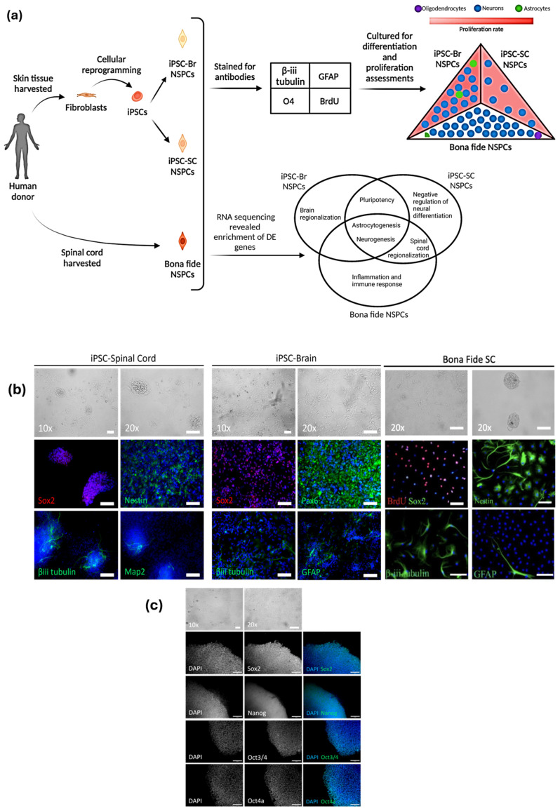
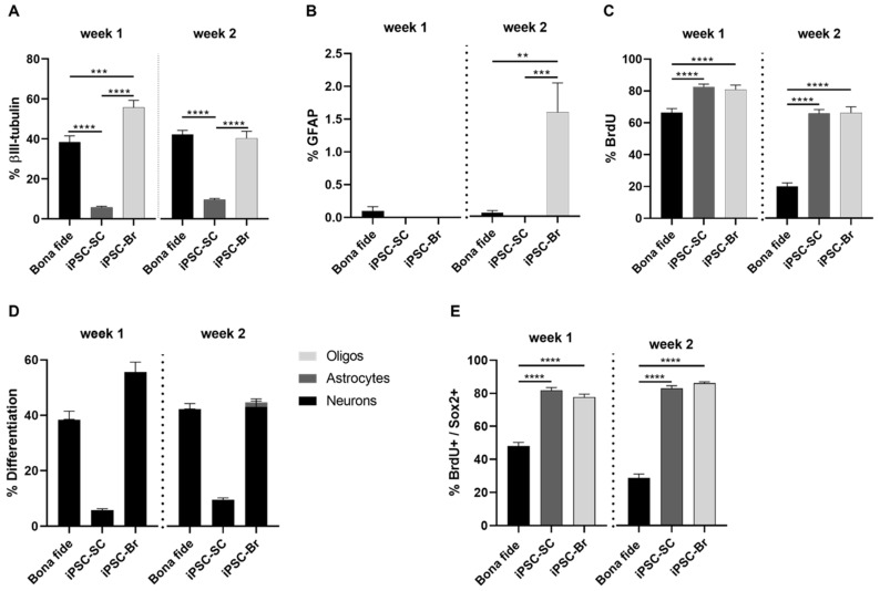
The adult human spinal cord harbors diverse populations of neural stem/progenitor cells (NSPCs) essential for neuroregeneration and central nervous system repair. While induced pluripotent stem cell (iPSC)-derived NSPCs offer significant therapeutic potential, understanding their molecular and functional alignment with bona fide spinal cord NSPCs is crucial for developing autologous cell therapies that enhance spinal cord regeneration and minimize immune rejection. In this study, we present the first direct transcriptomic and functional comparison of syngeneic adult human NSPC populations, including bona fide spinal cord NSPCs and iPSC-derived NSPCs regionalized to the spinal cord (iPSC-SC) and forebrain (iPSC-Br). RNA sequencing analysis revealed distinct transcriptomic profiles and functional disparities among NSPC types. iPSC-Br NSPCs exhibited a close resemblance to bona fide spinal cord NSPCs, characterized by enriched expression of neurogenesis, axon guidance, synaptic signaling, and voltage-gated calcium channel activity pathways. Conversely, iPSC-SC NSPCs displayed significant heterogeneity, suboptimal regional specification, and elevated expression of neural crest and immune response-associated genes. Functional assays corroborated the transcriptomic findings, demonstrating superior neurogenic potential in iPSC-Br NSPCs. Additionally, we assessed donor-specific influences on NSPC behavior by analyzing gene expression and differentiation outcomes across syngeneic populations from multiple individuals. Donor-specific factors significantly modulated transcriptomic profiles, with notable variability in the alignment of iPSC-derived NSPCs to bona fide spinal cord NSPCs. Enrichment of pathways related to neurogenesis, axon guidance, and synaptic signaling varied across donors, highlighting the impact of genetic and epigenetic individuality on NSPC behavior.

Keywords: cellular heterogeneity; differentiation potential; induced pluripotent stem cells; neural stem/progenitor cells; neurogenesis; patient-specific variability; proliferation dynamics; regenerative medicine; spinal cord injury; transcriptomics.


Conflict of interest statement

The authors declare no conflicts of interest.

Figures

Figure 1
Figure 1
Differentiation, proliferation, and characterization of NSPCs derived from iPSCs and bona fide source. iPSC-Br NSPCs more closely resemble bona fide NSPCs in morphology and marker expression compared to iPSC-SC NSPCs. (a) Schematic illustration of the experimental workflow. Skin tissue samples from human donors were cultured into primary fibroblasts and reprogrammed into iPSCs. (b) Morphological and immunocytochemical analysis of NSPCs. iPSC-SC NSPCs form flat and neurosphere-like colonies, expressing Sox2, Nestin, β-III tubulin, and Map2. iPSC-Br NSPCs exhibit multipolar spindle-like cells, expressing Sox2, Pax6, β-III tubulin, and GFAP. Bona fide spinal cord NSPCs form adherent layers and neurosphere-like colonies, expressing Nestin, Sox2, β-III tubulin, and GFAP. (c) Pluripotency markers (Sox2, Nanog, Oct3/4, Oct4a) in iPSC-SC colonies confirm retention of stemness. DAPI shows nuclei in all panels.
Figure 2
Figure 2
Comparative analysis of differentiation and proliferation profiles between bona fide and iPSC-derived NSPC. iPSC-Br NSPCs showed greater neuronal and astrocyte differentiation and higher proliferation rates than both iPSC-SC and bona fide NSPCs, indicating their superior neurogenic potential. (A) The percentage of β-III tubulin+ neurons was significantly higher in bona fide, and iPSC-Br NSPCs compared to iPSC-SC NSPCs at both time points. (B) By week two, GFAP+ astrocyte differentiation was markedly higher in iPSC-Br NSPCs compared to bona fide and iPSC-SC NSPCs, with minimal astrocytic differentiation observed in the latter. (C) Proliferation rates, measured as BrdU+ cell percentages, were significantly higher in iPSC-derived NSPCs compared to bona fide NSPCs under differentiation conditions. (D) Lineage differentiation profiles revealed that iPSC-Br NSPCs exhibited contributions from neuronal, astrocytic, and minimal oligodendrocytic lineages, while iPSC-SC NSPCs were predominantly neuronal. Bona fide NSPCs were also predominantly neuronal with negligible astrocytic and almost no oligodendrocytic differentiation. (E) Self-renewal capacity, as indicated by BrdU+/Sox2+ cells, was higher in iPSC-derived NSPCs compared to bona fide NSPCs at both time points. Data are presented as mean ± s.e.m.; ** p < 0.01, *** p < 0.001, **** p < 0.0001; n = 3 for bona fide, iPSC-SC, and iPSC-Br NSPCs. Each assay was conducted with three biological replicates (H17, H18, H25), and each functional assay, such as the percentage of β-III tubulin + cells, was performed with three technical replicates. Each technical replicate involved capturing 10 standardized images from a 96-well plate.
Figure 3
Figure 3
The transcriptomes of iPSC-Br and bona fide NSPCs are more similar than iPSC-SC and bona fide NSPCs. The gene expression analysis reveals that iPSC−Br NSPCs retain transcriptomic features similar to bona fide NSPCs, unlike the more distinct iPSC−SC NSPCs (A) Venn diagram of DE genes in both iPSC−SC and iPSC−Br NSPCs relative to bona fide NSPCs, including directionality (upregulation or downregulation). A total of 4600 genes were DE in both iPSC−SC and iPSC−Br NSPCs with a fold change ≥ 2 and q-value < 0.05. (B) The top 30 genes DE in the same direction (upregulation or downregulation) in both iPSC−SC and iPSC−Br NSPCs relative to bona fide NSPCs. (C) PCA of the 500 most variable genes across bona fide, iPSC-SC, and iPSC-Br NSPCs. Each dot represents a sample (H17, H18, H25), with donor-specific identifiers labeled. PC1 (69% variance) explains the largest transcriptomic differences, while PC2 (7% variance) and PC3 (6% variance) capture additional variation. iPSC-Br NSPCs cluster closer to bona fide NSPCs than iPSC-SC NSPCs, reflecting greater transcriptomic similarity. Donor-specific differences are evident along PC2, while PC3 highlights intrinsic differences between iPSC-SC and iPSC-Br NSPCs. All differences are statistically significant, * p < 0.05 for all comparisons. (D) Hierarchical clustering analysis for all transcripts (24,006 genes) found in common among bona fide, iPSC−SC, and iPSC−Br NSPCs. The clustering illustrates that bona fide NSPCs group closely together, with iPSC-Br NSPCs forming a cluster more similar to bona fide NSPCs than iPSC-SC NSPCs. The intensity of the green shading reflects expression levels, with darker shades indicating higher expression.
Figure 4
Figure 4
Comparative gene expression analysis of iPSC-derived NSPCs and bona fide NSPCs. iPSC-SC NSPCs may reflect a distinct reprogramming pathway compared to iPSC-Br NSPCs. (A) Volcano plots show differentially expressed (DE) genes between iPSC-SC NSPCs (left panel) and iPSC-Br NSPCs (right panel) relative to bona fide NSPCs, with red dots indicating significant DE genes (fold change ≥ 2, p-value < 0.05). (B) Heatmaps with hierarchical clustering depict gene expression patterns, comparing iPSC-SC NSPCs (left panel) and iPSC-Br NSPCs (right panel) to bona fide NSPCs. (C) Scatter plots highlight genes uniquely differentially expressed in iPSC-SC NSPCs (left panel) and iPSC-Br NSPCs (right panel) compared to bona fide NSPCs. (D) Lists of the top 50 upregulated and downregulated genes in iPSC-SC NSPCs and iPSC-Br NSPCs, each relative to bona fide NSPCs.
Figure 5
Figure 5
Comparative gene set enrichment in iPSC-derived NSPCs relative to bona fide NSPCs. Gene set enrichment analysis was conducted using the GSEA Preranked algorithm against a subset of MSigDB gene sets. A normalized enrichment score (NES) was used to compare gene sets across NSPCs in the categories of (A) hallmark gene sets, (B) GO molecular functions, (C) canonical pathways, (D) GO biological processes, and (E) cell-type signatures. Gene sets with significant positive or negative NES are indicated by *, representing a q-value < 0.05. iPSC-SC (n = 3) and iPSC-Brain (n = 3) are represented by pink and gray bars, respectively. Protein–protein interaction network maps of the top 30 (F) upregulated and (G) downregulated DE genes in both iPSC-SC and iPSC-Br NSPCs relative to bona fide NSPCs were generated using STRING v12.0 software (PPI enrichment p-value < 1 × 10⁻¹⁶). Each node represents proteins from a single, protein-coding gene locus, with colored nodes indicating query proteins and first-shell interactions, and white nodes representing second-shell interactions. Edge confidence is proportional to line thickness, indicating the strength of protein–protein associations.

Similar articles

Cited by

References

    1. Alizadeh A., Dyck S.M., Karimi-Abdolrezaee S. Traumatic Spinal Cord Injury: An Overview of Pathophysiology, Models and Acute Injury Mechanisms. Front. Neurol. 2019;10:282. doi: 10.3389/fneur.2019.00282. - DOI - PMC - PubMed
    1. Saremi J., Mahmoodi N., Rasouli M., Ranjbar F.E., Mazaheri E.L., Akbari M., Hasanzadeh E., Azami M. Advanced approaches to regenerate spinal cord injury: The development of cell and tissue engineering therapy and combinational treatments. Biomed. Pharmacother. 2022;146:112529. doi: 10.1016/j.biopha.2021.112529. - DOI - PubMed
    1. Yamazaki K., Kawabori M., Seki T., Houkin K. Clinical Trials of Stem Cell Treatment for Spinal Cord Injury. Int. J. Mol. Sci. 2020;21:3994. doi: 10.3390/ijms21113994. - DOI - PMC - PubMed
    1. Tetzlaff W., Okon E.B., Karimi-Abdolrezaee S., Hill C.E., Sparling J.S., Plemel J.R., Plunet W.T., Tsai E.C., Baptiste D., Smithson L.J., et al. A systematic review of cellular transplantation therapies for spinal cord injury. J. Neurotrauma. 2011;28:1611–1682. doi: 10.1089/neu.2009.1177. - DOI - PMC - PubMed
    1. Zeng C.-W. Advancing Spinal Cord Injury Treatment through Stem Cell Therapy: A Comprehensive Review of Cell Types, Challenges, and Emerging Technologies in Regenerative Medicine. Int. J. Mol. Sci. 2023;24:14349. doi: 10.3390/ijms241814349. - DOI - PMC - PubMed

Publication types

MeSH terms

LinkOut - more resources