. 2025 Jan 9;14(2):82.
doi: 10.3390/cells14020082.

Extracellular Vesicles Derived from Human Umbilical Mesenchymal Stem Cells Transfected with miR-7704 Improved Damaged Cartilage and Reduced Matrix Metallopeptidase 13

Affiliations

Extracellular Vesicles Derived from Human Umbilical Mesenchymal Stem Cells Transfected with miR-7704 Improved Damaged Cartilage and Reduced Matrix Metallopeptidase 13

Kun-Chi Wu et al. Cells. .

Abstract

We aimed to explore the therapeutic efficacy of miR-7704-modified extracellular vesicles (EVs) derived from human umbilical cord mesenchymal stem cells (HUCMSCs) for osteoarthritis (OA) treatment. In vitro experiments demonstrated the successful transfection of miR-7704 into HUCMSCs and the isolation of EVs from these cells. In vivo experiments used an OA mouse model to assess the effects of the injection of miR-7704-modified EVs intra-articularly. Walking capacity (rotarod test), cartilage morphology, histological scores, and the expression of type II collagen, aggrecan, interleukin-1 beta, and matrix metalloproteinase 13 (MMP13) in the cartilage were evaluated. The EVs were characterized to confirm their suitability for therapeutic use. IL-1beta-treated chondrocytes increased type II collagen and decreased MMP13 after treatment with miR-7704-overexpressed EVs. In vivo experiments revealed that an intra-articular injection of miR-7704-overexpressed EVs significantly improved walking capacity, preserved cartilage morphology, and resulted in higher histological scores compared to in the controls. Furthermore, the decreased expression of MMP13 in the cartilage post treatment suggests a potential mechanism for the observed therapeutic effects. Therefore, miR-7704-overexpressed EVs derived from HUCMSCs showed potential as an innovative therapeutic strategy for treating OA. Further investigations should focus on optimizing dosage, understanding mechanisms, ensuring safety and efficacy, developing advanced delivery systems, and conducting early-phase clinical trials to establish the therapeutic potential of HUCMSC-derived EVs for OA management.

Keywords: MMP13; extracellular vesicles; human umbilical cord mesenchymal stem cells; miRNA; osteoarthritis.


Conflict of interest statement

The authors declare no conflicts of interest.

Figures

Figure 1
Figure 1
Characteristics of HUCMSCs. (A) Surface marker expression of HUCMSCs. (BD) Adipogenesis. (B) Oil Red O staining of adipocytes differentiated from HUCMSCs. (C) Quantification of Oil Red staining at optical density (OD) 510. (D) Gene expressions of adipocyte-related genes (FABP4 and PPAR-γ). (EG) Osteogenesis. (E) Alizarin Red staining of HUCMSC-differentiated osteoblasts. (C) Quantification of Alizarin Red staining at OD 562. (D) Gene expressions of osteoblast-related genes (APAL and RUNX2). (H) Hematoxylin and eosin staining; immunohistochemistry of type II collagen and aggrecan of HUCMSC-differentiated chondrocytes. (I) Gene expressions of chondrocyte-related genes (ACAN and COL2A1). Scale bar = 100 μm. *** p < 0.001.
Figure 2
Figure 2
Characteristics of extracellular vesicles (EVs) derived from miR-7704-transfected HUCMSCs. (A) HUCMSCs transfected with GFP-tagged miR7704. GFP is revealed to be successfully transfected into HUCMSCs. (B) qPCR shows the gene expression of miR-7704 in the EVs derived from HUCMSC-, lenti-ctrl-, and miR-7704-transfected HUCMSCs. (C) Nanoparticle tracking analysis for measuring the size and concentration of EVs. (D) Transmission electron microscopy is used to observe the EVs; scale bar = 100 nm. (E) Western blot characterization of EVs’ marker proteins (CD9, 63, and 81). *** p < 0.001. Original images of (E) can be found in Supplementary Materials.
Figure 3
Figure 3
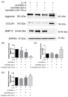
Western blot analysis of chondrocytes treated with IL-1beta 10ng/mL for 24 h, and then various extracellular vesicles (EVs) are treated. (A) Representative image of Western blotting. (B) Quantification of MMP13 (n = 3). (C) Quantification of type II collagen (COL2A1) (n = 3). (D) Quantification of aggrecan (n = 3). ** p < 0.01, *** p < 0.001. HUCMSCs: human umbilical cord mesenchymal stem cells; e: EVs; miR: microRNA. Original images of (A) can be found in Supplementary Materials.
Figure 4
Figure 4
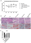
Rotarod performance test, histology, and The International Cartilage Repair Society (ICRS) score of osteoarthritis (OA) mice treated with HUCMSCs with or without transfection with lenti-ctrl or lenti-miR-7704 (n = 3 in each group). (A) Compared to the OA group, the OA mice that received HUCMSCs-EVs and lenti-miR-7704-EVs showed significant improvement after day 21. OA transplanted with lenti-ctrl EVs showed improvement on day 28. Ctrl: control, * p < 0.05. (B) The histology of the mouse OA model after 28 days of experiments (hematoxylin and eosin and Safranin O staining). The representative image of each group is illustrated. Scale bar = 100 μm. (C) The ICRS scores of the cartilage after various treatments. OA: osteoarthritis. A statistical test was performed using one-way ANOVA with a post hoc and Bonferroni test. * p < 0.05, ** p < 0.01.
Figure 5
Figure 5
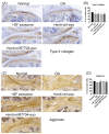
Immunohistochemical analysis of cartilage in mouse osteoarthritis (OA) model. (A) Type II collagen staining image from normal knee, OA, HUCMSCs-EVs, lenti-control-EVs, and miR-7704-EVs. (B) Quantitative analysis of the percentage of positively stained cells across five randomly selected fields (mean ± standard deviation). (C) Representative image of aggrecan for each group. (D) Quantitative analysis of the percentage of positively stained cells across five randomly selected fields (mean ± standard deviation).
Figure 6
Figure 6
Immunohistochemical analysis of cartilage in mouse osteoarthritis (OA) model. (A) IL-1β staining image from normal knee, OA, HUCMSCs-EVs, lenti-control-EVs, and lenti-miR-7704-EVs. (B) Quantitative analysis of the percentage of positively stained cells across five randomly selected fields (mean ± standard deviation). (C) Representative image of MMP13 for each group. (D) Quantitative analysis of the percentage of positively stained cells across five randomly selected fields (mean ± standard deviation). * p < 0.05, ** p < 0.01.

Similar articles

Cited by

References

    1. Chen X., Tang H., Lin J., Zeng R. Temporal Trends in the Disease Burden of Osteoarthritis from 1990 to 2019, and Projections until 2030. PLoS ONE. 2023;18:e0288561. doi: 10.1371/journal.pone.0288561. - DOI - PMC - PubMed
    1. Leifer V.P., Katz J.N., Losina E. The Burden of OA-Health Services and Economics. Osteoarthr. Cartil. 2022;30:10–16. doi: 10.1016/j.joca.2021.05.007. - DOI - PMC - PubMed
    1. Yahaya I., Wright T., Babatunde O.O., Corp N., Helliwell T., Dikomitis L., Mallen C.D. Prevalence of Osteoarthritis in Lower Middle- and Low-Income Countries: A Systematic Review and Meta-Analysis. Rheumatol. Int. 2021;41:1221–1231. doi: 10.1007/s00296-021-04838-y. - DOI - PMC - PubMed
    1. Li H., Kong W., Liang Y., Sun H. Burden of osteoarthritis in China, 1990–2019: Findings from the Global Burden of Disease Study 2019. Clin. Rheumatol. 2024;43:1189–1197. doi: 10.1007/s10067-024-06885-9. - DOI - PMC - PubMed
    1. Richard M.J., Driban J.B., McAlindon T.E. Pharmaceutical Treatment of Osteoarthritis. Osteoarthr. Cartil. 2023;31:458–466. doi: 10.1016/j.joca.2022.11.005. - DOI - PubMed

Publication types

MeSH terms

LinkOut - more resources