. 2025 Jan 13;14(2):108.
doi: 10.3390/cells14020108.

Blockade of TIPE2-Mediated Ferroptosis of Myeloid-Derived Suppressor Cells Achieves the Full Potential of Combinatory Ferroptosis and Anti-PD-L1 Cancer Immunotherapy

Affiliations

Blockade of TIPE2-Mediated Ferroptosis of Myeloid-Derived Suppressor Cells Achieves the Full Potential of Combinatory Ferroptosis and Anti-PD-L1 Cancer Immunotherapy

Hafiza Kashaf Tariq et al. Cells. .

Abstract

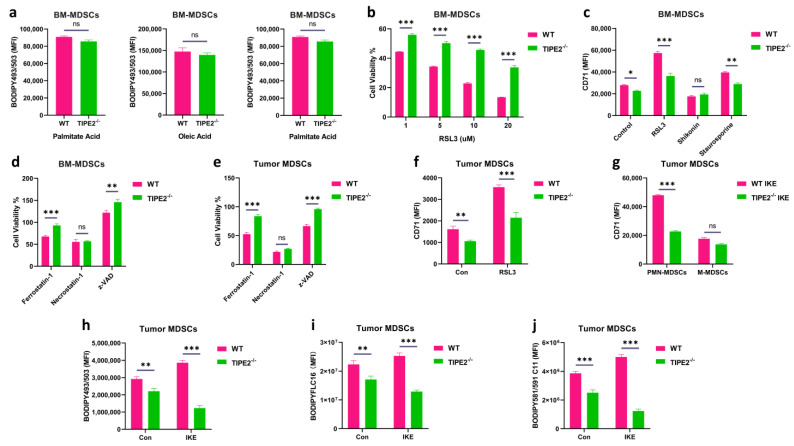
Although immune checkpoint blockade (ICB) therapy has attained unprecedented clinical success, the tolerance and immune suppression mechanisms evolved by tumor cells and their tumor microenvironment (TME) hinder its maximum anti-cancer potential. Ferroptosis therapy can partially improve the efficacy of ICB, but it is still subject to immune suppression by myeloid-derived suppressor cells (MDSCs) in the TME. Recent research suggests that an MDSC blockade can unleash the full therapeutic potential of the combined therapy of ferroptosis and ICB in liver cancer treatment. However, whether blocking the intrinsic ferroptosis pathways of MDSCs can relieve imidazole ketone erastin (IKE)-initiated ferroptosis-induced immune suppression and ultimately trigger the optimal therapeutic effect of the combined ferroptosis and ICB therapy is still unknown. Here, we report that TIPE2, a phospholipid transfer protein, regulated the ferroptosis susceptibility in MDSCs through reprogramming lipid peroxidation-related phosphatidylethanolamine (PE) and phosphatidylcholine (PC) species composition. TIPE2-deficient MDSCs resisted IKE-induced ferroptosis by up-regulating SLC7A11 and GPX4, and dissolved ferroptosis-induced immunosuppressive function by down-regulating lipid ROS whilst encouraging T cell proliferation and infiltration into tumor tissues to improve ferroptosis therapy. More importantly, TIPE2-deficient MDSCs achieved the full anti-tumor therapeutic potential of IKE-induced ferroptosis therapy and a PD-L1 blockade. These findings indicate that TIPE2 confers the ferroptosis sensitivity of MDSCs, and combining the targeting of the TIPE2 of MDSCs, ferroptosis therapy, and ICB is a novel therapeutic option for cancer treatment.

Keywords: MDSCs; TIPE2; cancer treatment; ferroptosis; immune checkpoint blockade.


Conflict of interest statement

The authors declare no conflicts of interest.

Figures

Figure 1
Figure 1
TIPE2 is involved in the induction of ferroptosis in MDSCs. (a) Fatty acid accumulation with lipophilic fluorescent dye BODIPY493/503 in Gr−1+ MDSCs extracted from the bone marrow treated with palmitate acid (saturated fatty acid, 100 μM), linoleic acid (polyunsaturated fatty acid, 100 μM), and oleic acid (monounsaturated fatty acid, 100 μM) for 48 h. Mean ± SD are shown. (b) AlamarBlue cell viability assay on Gr−1+ MDSCs extracted from the bone marrow incubated with different concentrations 1 µM, 5 µM, 10 µM, and 20 µM of RSL3 for 24 h. Mean ± SD are shown. (c) CD71 expression in Gr−1+ MDSCs extracted from the bone marrow in the presence of different cell death inducers for 18 h: ferroptosis (RSL3, 20 µM), necroptosis (Shikonin, 1 µM), and apoptosis (Staurosporine, 0.25 µM). Mean ± SD are shown. (d) AlamarBlue cell viability assay on Gr-1+ MDSCs extracted from the bone marrow in the presence of different cell death inhibitors for 24 h: 1 µM ferrostatin-1 for ferroptosis, 1 µM necrostatin−1 for necroptosis, and 10 µM z−VAD−FMK (zVAD) for apoptosis. Mean ± SD are shown. (e) AlamarBlue cell viability assay on Gr−1+ MDSCs extracted from the LLC TB mice in the presence of different cell death inhibitors for 24 h: 1 µM ferrostatin-1 for ferroptosis, 1 µM necrostatin-1 for necroptosis, and 10 µM z−VAD−FMK (zVAD) for apoptosis. Mean ± SD are shown. (f) CD71 expression in Gr−1+ MDSCs extracted from the LLC TB mice in the presence of ferroptosis inducer 10 µM RSL3 for 24 h. Mean ± SD are shown. (g) CD71 expression in PMN−MDSCs and M−MDSCs extracted from the LLC TB mice. Mean ± SD are shown. (h) Lipid level with lipophilic fluorescent dye BODIPY493/503 in Gr−1+ MDSCs extracted from the LLC TB mice treated with IKE. Mean ± SD are shown. (i) Lipid uptake with palmitic acid fluorescent dye BODIPYFLC16 in Gr−1+ MDSCs extracted from the LLC TB mice treated with IKE. Mean ± SD are shown. (j) Lipid peroxidation level with fluorescent dye BODIPY 581/591 C11 in Gr−1+ MDSCs extracted from the LLC TB mice treated with IKE. Mean ± SD are shown. (aj) Data are expressed as *, p ≤ 0.05, **, p ≤ 0.01, ***, p ≤ 0.001, or ns, no significant difference.
Figure 2
Figure 2
TIPE2 regulates ferroptosis-related genes of MDSCs. (a) mRNA expression of ferroptosis-promoting genes in MDSCs. qRT−PCR was conducted in Gr−1+ MDSCs extracted from the bone marrow and treated with RSL3. Mean ± SD are shown. (b) mRNA expression of ferroptosis resistance genes in MDSCs. qRT−PCR was conducted in Gr−1+ MDSCs extracted from the bone marrow and treated with RSL3. Mean ± SD are shown. (c) mRNA expression of ferroptosis−promoting genes in MDSCs. qRT-PCR was conducted in Gr−1+ MDSCs extracted from the LLC TB mice treated with IKE. Mean ± SD are shown. (d) mRNA expression of ferroptosis resistance genes in MDSCs. qRT−PCR was conducted in Gr−1+ MDSCs extracted from the LLC TB mice treated with IKE. Mean ± SD are shown. (e) Protein expression of ferroptosis−promoting and resistance genes in MDSCs. Western blot was conducted in Gr−1+ MDSCs extracted from the LLC TB mice treated with IKE. Mean ± SD are shown. (ad) Data are expressed as ***, p ≤ 0.001, or ns, no significant difference.
Figure 3
Figure 3
TIPE2−deficient MDSCs decrease ferroptosis−induced suppressive function. (a) ROS level using the CM−H2DCFDA fluorescent dye in Gr−1+ extracted from the LLC TB mice treated with IKE. Mean ± SD are shown. (b) NO level using the DAF−FM DA fluorescent dye in Gr−1+ MDSCs extracted from the LLC TB mice treated with IKE. Mean ± SD are shown. (c) mRNA expression of Arg1 and Inos in MDSCs. qRT−PCR conducted in Gr−1+ MDSCs extracted from the LLC TB mice treated with IKE. Mean ± SD are shown. (d) Protein expression of ARG1 and iNOS in MDSCs. Western blot conducted in Gr−1+ MDSCs extracted from the LLC TB mice treated with IKE. Mean ± SD are shown. (e) CD71 expression in Gr-1+ extracted from the LLC TB mice treated with ROS inhibitor (L−NAC, 1 mM). Mean ± SD are shown. (f) The expression of T cell proliferation in Gr−1+ MDSCs extracted from the LLC TB mice treated with 40 µM IKE (ferroptosis inducer) for 6 h, washed extensively, and cocultured with CFSE−labeled CD3+ T cells at the ratio of 1:2 and 1:4. Mean ± SD are shown. (g) The expression of T cell proliferation in Gr−1+ MDSCs extracted from the LLC TB mice treated with ROS inhibitor (L−NAC, 1 mM) and cocultured with CFSE−labeled CD3+ T cells at the ratio of 1:1. Mean ± SD are shown. (ac,eg) Data are expressed as **, p ≤ 0.01, or ***, p ≤ 0.001.
Figure 4
Figure 4
TIPE2-deficient MDSCs enhance ferroptosis-induced tumor growth inhibition via reprogramming the immune microenvironment. (a) A schematic representation of the experimental design; C57BL/6 mice were injected s.c. with LLC cells on day 0. Then, IKE (10mg/kg) was injected i.p. at day 8 till day 21. (b) The tumor volume of LLC C57BL/6 mice (n = 3 mice/group) treated with IKE for 2 weeks. Mean ± SD are shown. (c) The tumor weight of LLC C57BL/6 mice (n = 3 mice/group) treated with IKE for 2 weeks. Mean ± SD are shown. (d) The body weight of LLC C57BL/6 mice (n = 3 mice/group) treated with IKE for 2 weeks. Mean ± SD are shown. (e) A schematic representation of the experimental design, C57BL/6 mice were injected s.c. with B16F10 cells on day 0. Then, IKE (10 mg/kg) was injected i.p. at day 8 till day 21. (f) The tumor volume of B16F10 C57BL/6 mice (n = 3 mice/group) treated with IKE for 2 weeks. Mean ± SD are shown. (g) The tumor weight of B16F10 C57BL/6 mice (n = 3 mice/group) treated with IKE for 2 weeks. Mean ± SD are shown. (h) The body weight of B16F10 C57BL/6 mice (n = 3 mice/group) treated with IKE for 2 weeks. Mean ± SD are shown. (i) The percentages of immune cells in tumor tissues extracted from the LLC TB mice treated with IKE: MDSCs, PMN-MDSCs, M-MDSCs, Treg cells, DCs, macrophages, B cells, NK cells, CD3+ T cells, CD4+ T cells, CD8+ T cells, were discovered by flow cytometry. MDSCs were CD45+ CD11b+ Gr-1+ cells; PMN-MDSCs were CD45+ CD11b+ Ly6CLy6G+ cells; M-MDSCs were CD45+ CD11b+ Ly6C+ Ly6G cells; Treg cells were CD45+ CD3+ CD4+ CD25+ CD127 cells; DCs were CD45+ CD11b+ CD11c+ cells; macrophages were CD45+CD11b+ F4/80+ cells; B cells were CD45+CD3 CD19+ cells; and NK cells were CD45+ CD3 NK1.1+ cells; CD3+ T cells were CD45+ CD3+ cells; CD4+ T cells were CD45+ CD3+CD4+ CD8 cells; CD8+ T cells were CD45+CD3+CD4CD8+ cells. Mean ± SD are shown. (bd,fi) Data are expressed as ***, p ≤ 0.001, or ns, no significant difference.
Figure 5
Figure 5
The ferroptosis therapy of TIPE2−deficient MDSCs enhances anti−PD−L1 cancer immunotherapy. (a) A schematic representation of the experimental design C57BL/6 mice were injected s.c. with LLC cells on day 0. Then, IKE (10mg/kg) was injected i.p. at day 8 till day 21. Also, anti−PD−L1 (200 μg) was injected i.p. every 3 days. (b) The tumor volume of LLC C57BL/6 mice (n = 3 mice/group) treated with IKE for 2 weeks. Mean ± SD are shown. (c) The tumor weight of LLC C57BL/6 mice (n = 3 mice/group) treated with IKE for 2 weeks. Mean ± SD are shown. (d) The body weight of LLC C57BL/6 mice (n = 3 mice/group) treated with IKE for 2 weeks. Mean ± SD are shown. (e) A schematic representation of the experimental design C57BL/6 mice were injected s.c. with B16F10 cells on day 0. Then, IKE (10mg/kg) was injected i.p. at day 8 till day 21. Also, anti−PD−L1 (200 μg) was injected i.p. every 3 days. (f) The tumor volume of B16F10 C57BL/6 mice (n = 3 mice/group) treated with IKE for 2 weeks. Mean ± SD are shown. (g) The tumor weight of B16F10 C57BL/6 mice (n = 3 mice/group) treated with IKE for 2 weeks. Mean ± SD are shown. (h) The body weight of B16F10 C57BL/6 mice (n = 3 mice/group) treated with IKE for 2 weeks. Mean ± SD are shown. (i) The percentages of immune cells in tumor tissues extracted from the LLC TB mice treated with IKE: MDSCs, PMN−MDSCs, M−MDSCs, Treg cells, DCs, macrophages, B cells, NK cells, CD3+ T cells, CD4+ T cells, CD8+ T cells, were discovered by flow cytometry. MDSCs were CD45+ CD11b+ Gr−1+ cells; PMN−MDSCs were CD45+ CD11b+ Ly6CLy6G+ cells; M−MDSCs were CD45+ CD11b+ Ly6C+ Ly6G cells; Treg cells were CD45+ CD3+ CD4+ CD25+ CD127 cells; DCs were CD45+ CD11b+ CD11c+ cells; macrophages were CD45+CD11b+ F4/80+ cells; B cells were CD45+CD3 CD19+ cells; and NK cells were CD45+ CD3 NK1.1+ cells; CD3+ T cells were CD45+ CD3+ cells; CD4+ T cells were CD45+ CD3+CD4+ CD8 cells; CD8+ T cells were CD45+CD3+CD4CD8+ cells. Mean ± SD are shown. (bd,fi) Data are expressed as ***, p ≤ 0.001, or ns, no significant difference.

References

    1. Sharma P., Siddiqui B.A., Anandhan S., Yadav S.S., Subudhi S.K., Gao J., Goswami S., Allison J.P. The Next Decade of Immune Checkpoint Therapy. Cancer Discov. 2021;11:838–857. doi: 10.1158/2159-8290.CD-20-1680. - DOI - PubMed
    1. Huang A.C., Zappasodi R. A Decade of Checkpoint Blockade Immunotherapy in Melanoma: Understanding the Molecular Basis for Immune Sensitivity and Resistance. Nat. Immunol. 2022;23:660–670. doi: 10.1038/s41590-022-01141-1. - DOI - PMC - PubMed
    1. Morad G., Helmink B.A., Sharma P., Wargo J.A. Hallmarks of Response, Resistance, and Toxicity to Immune Checkpoint Blockade. Cell. 2021;184:5309–5337. doi: 10.1016/j.cell.2021.09.020. - DOI - PMC - PubMed
    1. Haddad A.F., Young J.S., Gill S., Aghi M.K. Resistance to Immune Checkpoint Blockade: Mechanisms, Counter-Acting Approaches, and Future Directions. Semin. Cancer Biol. 2022;86:532–541. doi: 10.1016/j.semcancer.2022.02.019. - DOI - PMC - PubMed
    1. Soula M., Weber R.A., Zilka O., Alwaseem H., La K., Yen F., Molina H., Garcia-Bermudez J., Pratt D.A., Birsoy K. Metabolic Determinants of Cancer Cell Sensitivity to Canonical Ferroptosis Inducers. Nat. Chem. Biol. 2020;16:1351–1360. doi: 10.1038/s41589-020-0613-y. - DOI - PMC - PubMed

Publication types

MeSH terms

LinkOut - more resources