Review
. 2024 Dec 28;32(1):16.
doi: 10.3390/curroncol32010016.

Understanding the Immune System and Biospecimen-Based Response in Glioblastoma: A Practical Guide to Utilizing Signal Redundancy for Biomarker and Immune Signature Discovery

Affiliations
Review

Understanding the Immune System and Biospecimen-Based Response in Glioblastoma: A Practical Guide to Utilizing Signal Redundancy for Biomarker and Immune Signature Discovery

Luke R Jackson et al. Curr Oncol. .

Abstract

Glioblastoma (GBM) is a primary central nervous system malignancy with a median survival of 15-20 months. The presence of both intra- and intertumoral heterogeneity limits understanding of biological mechanisms leading to tumor resistance, including immune escape. An attractive field of research to examine treatment resistance are immune signatures composed of cluster of differentiation (CD) markers and cytokines. CD markers are surface markers expressed on various cells throughout the body, often associated with immune cells. Cytokines are the effector molecules of the immune system. Together, CD markers and cytokines can serve as useful biomarkers to reflect immune status in patients with GBM. However, there are gaps in the understanding of the intricate interactions between GBM and the peripheral immune system and how these interactions change with standard and immune-modulating treatments. The key to understanding the true nature of these interactions is through multi-omic analysis of tumor progression and treatment response. This review aims to identify potential non-invasive blood-based biomarkers that can contribute to an immune signature through multi-omic approaches, leading to a better understanding of immune involvement in GBM.

Keywords: biomarkers; glioblastoma; immune response; immune system; tumor resistance.


Conflict of interest statement

The authors declare no conflicts of interest.

Figures

Figure 3
Figure 3
(A) Immune cell markers, clusters of differentiation (CD), and (B) cytokine markers illustrating expression in healthy brain tissue as compared to blood based on highest expression in the brain and concentration in the blood, respectively. Average blood concentration values (left Y-axis) were plotted next to the brain tissue-specific maximum transcript levels (right Y-axis) for each marker with data obtained from the Human Protein Atlas (HPA) [82] showing the differential expression of blood and tissues. The Human Protein Atlas data for both blood and brain tissue specimens are representative of patients without GBM to help identify which immune blood-based biomarkers may be able to reflect changes in brain tissue. The Human Protein Atlas. Available online: https://www.proteinatlas.org/ (accessed 23 September 2024).
Figure 1
Figure 1
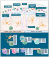
The brain as a site of active and passive immunity illustrating known linkages to cell types and their immune functions. Immune cells in the brain and their location (top panel), legend of immune cell types, and their respective markers (lower panel). While other immune cells, like mast and plasma cells, are known to play a role in CNS immune surveillance, there is limited data available demonstrating involvement in glioma, and their cell makers are redundant with the other cells listed here. Immune maker signatures were based on data established in previously published reviews [25,26,27,28,29,30,31]. Illustrations in this figure were created with Biorender.com (accessed on 23 September 2024).
Figure 2
Figure 2
Glioblastoma development leads to ambivalent alteration in immune function with ensuing evolution of immune marker profiles as consequences of tumor progression, biological aggressiveness, and subsequent management. These are reflected in immune signatures of varying degrees in tissue, CSF, and blood. Illustrations in this figure were created with Biorender.com and PowerPoint.
Figure 4
Figure 4
Overview of neuro-inflammation pathways. (A). Prominent signals associated with pro-inflammatory signaling (CCL2, IL6, TNF, IL10, and IL4) are illustrated downstream from NF-κβ in neuroinflammation pathways as identified in microglia. (B). Signals present in astrocytes in neuroinflammation leading to neurogenesis, Treg and T cell recruitment, microglial activation (adapted from Ingenuity Pathway Analysis (IPA)) [95].
Figure 5
Figure 5
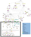
Overview of molecular pathways and immune signatures involved in PMT. Tumor cells produce cytokines, growth factors, and other relevant biomarkers (yellow) that increase invasive properties and have immunomodulating capacity, encouraging PMT. Effects of tumor immunomodulation (purple) and their impacts on tumor phenotypic behavior (peach) are linked above as well. Additional biomarkers are shown in the figure as transmembrane receptors and channels (green) and metabolites (blue) with their overall connections to immunomodulation and development of PMT (adapted from Ingenuity Pathway Analysis (IPA)) [95].
Figure 6
Figure 6
PD-1 immunotherapy illustrating dendritic cell markers (yellow) and the interplay between M2 macrophages, T cells, and tumor cells highlighting significant signaling pathways in GBM (magenta) in tumor cells and activated CD4+ and CD8+ T lymphocytes. Figure adapted from Ingenuity Pathway Analysis (IPA) [95].
Figure 7
Figure 7
Approach to the immune signal redundancy problem in GBM. Signal redundancy is multifactorial and evolves in stages from the normal CNS to GBM development/progression/management (left to right), and with radiation and chemotherapy to encompass a balance of immune suppression, evasion, and response as well as immune system exhaustion (upper panel, left to right). The redundancy cannot be modified; thus, emphasis is placed on enhanced utilization of clinically available data (step 1), linkage of multi-channel data across all types of biospecimens (step 2), and comparison with the normal CNS. Computational approaches can then be employed to normalize data and select the most important clinically relevant features (step 3), followed by validation aimed at the most promising signals and the use of novel therapies (step 4).

Similar articles

Cited by

References

    1. Stupp R., Mason W.P., van den Bent M.J., Weller M., Fisher B., Taphoorn M.J., Belanger K., Brandes A.A., Marosi C., Bogdahn U., et al. Radiotherapy plus concomitant and adjuvant temozolomide for glioblastoma. N. Engl. J. Med. 2005;352:987–996. doi: 10.1056/NEJMoa043330. - DOI - PubMed
    1. Stupp R., Hegi M.E., Mason W.P., van den Bent M.J., Taphoorn M.J., Janzer R.C., Ludwin S.K., Allgeier A., Fisher B., Belanger K., et al. Effects of radiotherapy with concomitant and adjuvant temozolomide versus radiotherapy alone on survival in glioblastoma in a randomised phase III study: 5-year analysis of the EORTC-NCIC trial. Lancet Oncol. 2009;10:459–466. doi: 10.1016/S1470-2045(09)70025-7. - DOI - PubMed
    1. Vaubel R.A., Tian S., Remonde D., Schroeder M.A., Mladek A.C., Kitange G.J., Caron A., Kollmeyer T.M., Grove R., Peng S., et al. Genomic and Phenotypic Characterization of a Broad Panel of Patient-Derived Xenografts Reflects the Diversity of Glioblastoma. Clin. Cancer Res. 2020;26:1094–1104. doi: 10.1158/1078-0432.CCR-19-0909. - DOI - PMC - PubMed
    1. Yabo Y.A., Heiland D.H. Understanding glioblastoma at the single-cell level: Recent advances and future challenges. PLoS Biol. 2024;22:e3002640. doi: 10.1371/journal.pbio.3002640. - DOI - PMC - PubMed
    1. Hanahan D. Hallmarks of Cancer: New Dimensions. Cancer Discov. 2022;12:31–46. doi: 10.1158/2159-8290.CD-21-1059. - DOI - PubMed

LinkOut - more resources