. 2025 Jan 4;11(1):36.
doi: 10.3390/gels11010036.

Polymer Gels Based on PAMAM Dendrimers Functionalized with Caffeic Acid for Wound-Healing Applications

Affiliations

Polymer Gels Based on PAMAM Dendrimers Functionalized with Caffeic Acid for Wound-Healing Applications

Ricardo I Castro et al. Gels. .

Abstract

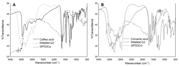
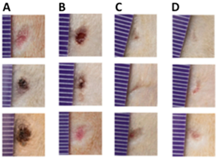
The wound-healing process has usually been related to therapeutic agents with antioxidant properties. Among them, caffeic acid, a cinnamic acid derivative, stands out. However, the use of this natural product is affected by its bioavailability and half-life. Nowadays, different approaches are being taken to improve the above-mentioned characteristics, as many active surface groups are present in polyamidoamine (PAMAM) dendrimers; without the need for extra cross-linking agents, physical gels are created by interactions such as hydrogen bonds, van der Waals forces, or π-π interactions based on the modification of the surface. One of these is functionalization with dendrimers, such as the poly(amidoamine) (PAMAM) family. To evaluate the effectiveness of functionalizing caffeic acid with PAMAM dendrimers, the in vitro and in vivo wound-healing properties of gel-PAMAM G3 conjugated with caffeic acid (GPG3Ca) and its precursor, cinnamic acid (GPG3Cin), were studied. The results showed no cytotoxicity and wound-healing activity at a concentration of 20 μg/mL in HaCaT cells with the GPG3Ca. Additionally, the ability to activate molecular mediators of the healing process was evidenced. Furthermore, GPG3Ca potentiated the in vivo wound-healing process. The positive effects and lack of cytotoxicity at the used concentration of the synthesized GPG3Ca on the wound-healing process could position it as an effective agent for wound-healing treatment.

Keywords: assay in vitro; caffeic and cinnamic acid; gel-PAMAM dendrimer; synthesis polymers gels; wound healing.


Conflict of interest statement

The authors declare that they have no known competing financial interests or personal relationships that could have appeared to influence the work reported in this paper.

Figures

Figure 1
Figure 1
Synthesis of GPG3Ca and GPG3Cin in the reaction formation of amides.
Figure 2
Figure 2
MALDI-TOF spectra of (A) GPG3Ca; (B) GPG3Cin; (C) PAMAM G3.
Figure 3
Figure 3
UV–visible spectra of (A) cinnamic acid; (B), caffeic acid; (C), GPG3Cin; (D) GPG3Ca.
Figure 4
Figure 4
FTIR spectra of (A) caffeic acid: solid line, PAMAN G3: dotted line, and GPG3Ca: dashed line; (B) cinnamic acid: solid line, PAMAN G3: dotted line, and GPG3Cin: dashed line.
Figure 5
Figure 5
Thermograms of GPG3Ca, GPG3Cin, and PAMAM G3 at temperatures between 50 and 700 °C.
Figure 6
Figure 6
HPLC chromatogram of (A) GPG3Ca; (B) GPG3Cin; (C) PAMAM G3.
Figure 7
Figure 7
HaCaT Cell viability at a concentration of 20 μg/mL of the different molecules studied. ** p < 0.01.
Figure 8
Figure 8
In vitro scratch assay. (A) Wound-closure percentages at 24 and 48 h. (B) Percentage of change between 24 and 48 h. *** p < 0.001 compared to GPG3Cin and the positive control; ++ p < 0.01 compared to the positive control; ### p < 0.001 compared to PAMAM G3, GPG3Cin, cinnamic and caffeic acid, and negative control; +++ p < 0.001 compared to the rest of the molecules.
Figure 9
Figure 9
Relative expression of (A) Ki67; (B) Cyclin D1; (C) IL-8; (D) MMP 1; (E) KRT 17. ** p < 0.01 and *** p < 0.001 compared to the basal expression.
Figure 10
Figure 10
Representative images of the in vivo wound-healing process (day 9). The columns represent (A) saline control group; (B) base cream group; (C) GPG3Ca 0.05% group; (D) GPG3Ca 0.1% group.
Figure 11
Figure 11
(A) Wound-healing process (9 days) of the different groups studied and (B) increase (folds) in wound closure at the end of the assay. * p < 0.05 compared to saline and base cream controls.
Figure 12
Figure 12
Histopathological analysis at day 9 of base cream group (A,B) and GPG3Ca 0.1% group (C,D). In (A,B), a partially recovered epidermis was observed (black arrow, image (B)); also, granulation tissue, neovascularization, and thin collagen fibers in a blue tone with Masson trichrome staining were observed in the dermis (white arrow, image (B)). In image (C,D), a completely re-epithelialized epidermis was observed (black arrow, image (D)); in addition, in the dermis, thicker collagen fibers with Masson trichrome staining and little granulation tissue were observed (white arrow, image (D)). Magnification (40×), scale bar 500 µm.

Similar articles

Cited by

References

    1. Yates C.C., Hebda P., Wells A. Skin wound healing and scarring: Fetal wounds and regenerative restitution. Birth Defects Res. Part C Embryo Today Rev. 2012;96:325–333. doi: 10.1002/bdrc.21024. - DOI - PMC - PubMed
    1. Biswas T.K., Pandit S., Chakrabarti S., Banerjee S., Poyra N., Seal T. Evaluation of Cynodon dactylon for wound healing activity. J. Ethnopharmacol. 2017;197:128–137. doi: 10.1016/j.jep.2016.07.065. - DOI - PubMed
    1. Lindley L.E., Stojadinovic O., Pastar I., Tomic-Canic M. Biology and biomarkers for wound healing. Plast. Reconstr. Surg. 2016;138:18S–28S. doi: 10.1097/PRS.0000000000002682. - DOI - PMC - PubMed
    1. Richardson M. Acute wounds: An overview of the physiological healing process. Nurs. Times. 2004;100:50–53. - PubMed
    1. Young A., McNaught C.-E. The physiology of wound healing. Surg. 2011;29:475–479. doi: 10.1016/j.mpsur.2011.06.011. - DOI

LinkOut - more resources