. 2025 Jan 4;11(1):37.
doi: 10.3390/gels11010037.

Kombucha Versus Vegetal Cellulose for Affordable Mucoadhesive (nano)Formulations

Affiliations

Kombucha Versus Vegetal Cellulose for Affordable Mucoadhesive (nano)Formulations

Ioana Popa-Tudor et al. Gels. .

Abstract

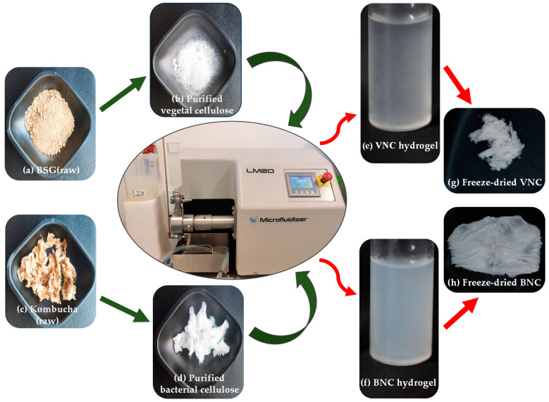
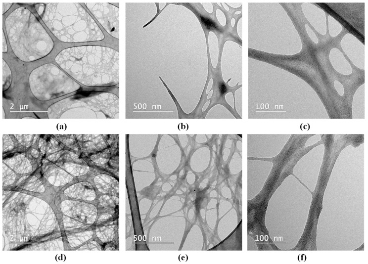
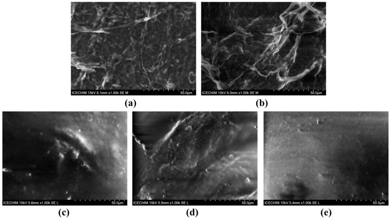
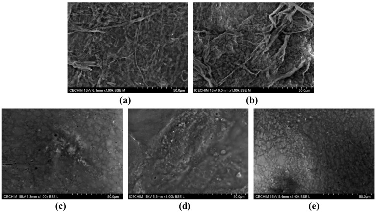
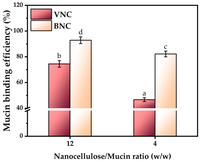
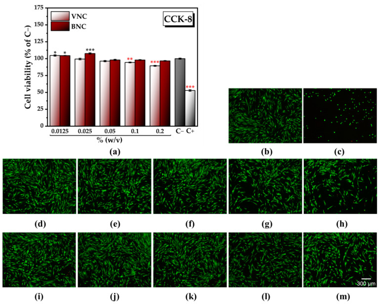
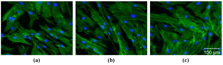
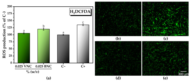
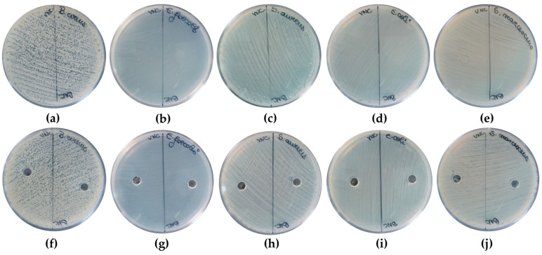
Cellulose nanofibers gained increasing interest in the production of medical devices such as mucoadhesive nanohydrogels due to their ability to retain moisture (high hydrophilicity), flexibility, superior porosity and durability, biodegradability, non-toxicity, and biocompatibility. In this work, we aimed to compare the suitability of selected bacterial and vegetal nanocellulose to form hydrogels for biomedical applications. The vegetal and bacterial cellulose nanofibers were synthesized from brewer's spent grains (BSG) and kombucha membranes, respectively. Two hydrogels were prepared, one based on the vegetal and the other based on the bacterial cellulose nanofibers (VNC and BNC, respectively). VNC was less opaque and more fluid than BNC. The cytocompatibility and in vitro antioxidant activity of the nanocellulose-based hydrogels were investigated using human gingival fibroblasts (HGF-1, ATCC CRL-2014). The investigation of the hydrogel-mucin interaction revealed that the BNC hydrogel had an approx. 2× higher mucin binding efficiency than the VNC hydrogel at a hydrogel/mucin ratio (mg/mg) = 4. The BNC hydrogel exhibited the highest potential to increase the number of metabolically active viable cells (107.60 ± 0.98% of cytotoxicity negative control) among all culture conditions. VNC reduced the amount of reactive oxygen species (ROS) by about 23% (105.5 ± 2.2% of C-) in comparison with the positive control, whereas the ROS level was slightly higher (120.2 ± 3.9% of C-) following the BNC hydrogel treatment. Neither of the two hydrogels showed antibacterial activity when assessed by the diffusion method. The data suggest that the BNC hydrogel based on nanocellulose from kombucha fermentation could be a better candidate for cytocompatible and mucoadhesive nanoformulations than the VNC hydrogel based on nanocellulose from brewer's spent grains. The antioxidant and antibacterial activity of BNC and both BNC and VNC, respectively, should be improved.

Keywords: brewer’s spent grains; cytocompatibility; gingival fibroblasts; hydrogels; kombucha fermentation; mucoadhesion; nanocellulose.


Conflict of interest statement

The authors declare no conflicts of interest.

Figures

Figure 1
Figure 1
Schematic representation of cellulose purification and hydrogel preparation: (a) brewer’s spent grains (BSG); (b) purified vegetal cellulose from BSG; (c) bacterial cellulose (BC) membrane from kombucha fermentation; (d) purified BC; (e) hydrogel of vegetal nanocellulose from BSG (VNC); (f) hydrogel of bacterial nanocellulose from kombucha fermentation (BNC); (g) freeze-dried VNC; and (h) freeze-dried BNC.
Figure 2
Figure 2
TEM analysis of: (a) VNC (2 µm scale); (b) VNC (500 nm scale), (c) VNC (100 nm scale); (d) BNC (2 µm scale); (e) BNC (500 nm scale); and (f) BNC (100 nm scale); VNC—hydrogel of vegetal nanocellulose from brewer’s spent grains; and BNC—hydrogel of bacterial nanocellulose from kombucha fermentation.
Figure 3
Figure 3
SEM analysis using secondary electrons (SE) detector (1000×) of: (a) VNC, (b) BNC, (c) VNCMu, (d) BNCMu, and (e) Mu; VNC—hydrogel of vegetal nanocellulose from brewer’s spent grains-based hydrogel; BNC—hydrogel of bacterial nanocellulose from kombucha fermentation-based hydrogel; VNCMu—VNC mixed with a 3.5% mucin suspension in a ratio of 1:1 (v/v); BNCMu—BNC mixed with a 3.5% mucin suspension in a ratio of 1:1 (v/v); and Mu—mucin suspension.
Figure 4
Figure 4
SEM analysis using backscattered electrons (BSE) detector (1000×) of: (a) VNC, (b) BNC, (c) VNCMu, (d) BNCMu, (e) Mu; VNC—hydrogel of vegetal nanocellulose from brewer’s spent grains; BNC—hydrogel of bacterial nanocellulose from kombucha fermentation; VNCMu—VNC mixed with a 3.5% mucin suspension in a ratio of 1:1 (v/v); BNCMu—BNC mixed with a 3.5% mucin suspension in a ratio of 1:1 (v/v); and Mu—mucin suspension.
Figure 5
Figure 5
X-ray diffraction (XRD) analysis and crystallinity index (Xc,%) of: (a) VNC, Mu, VNCMu; (b) BNC, Mu, BNCMu; the vertical bars represent the main diffraction peaks of cellulose Iα, Iβ, and amorphous cellulose in the PDXL database; VNC—hydrogel of vegetal nanocellulose from brewer’s spent grains; BNC—hydrogel of bacterial nanocellulose from kombucha fermentation; VNCMu—VNC mixed with a 3.5% mucin suspension in a ratio of 1:1 (v/v); BNCMu—BNC mixed with a 3.5% mucin suspension in a ratio of 1:1 (v/v); and Mu—mucin suspension.
Figure 6
Figure 6
Overlapping ATR-FTIR spectra for (a) VNC, Mu, VNCMu, and (b) BNC, Mu, and BNCMu. VNCMu—hydrogel of vegetal nanocellulose from brewer’s spent grains (VNC) mixed with a mucin suspension in a ratio of 1:1 (v/v); and BNCMu—hydrogel of bacterial nanocellulose from kombucha fermentation (BNC) mixed with a mucin suspension in a ratio of 1:1 (v/v).
Figure 7
Figure 7
Mucin binding efficiency (±standard deviation, n = 3, α < 0.05; different letters indicate statistically significant differences between samples); VNC—hydrogel of vegetal nanocellulose from brewer’s spent grains; and BNC—hydrogel of bacterial nanocellulose from kombucha fermentation.
Figure 8
Figure 8
Rheological behavior of VNC and BNC hydrogels: (a) frequency sweep of VNC; (b) frequency sweep of BNC; (c) flow sweep of VNC; (d) flow sweep of BNC; (e) axial mode of VNC; (f) axial mode of BNC; VNC—hydrogel of vegetal nanocellulose from brewer’s spent grains; and BNC—hydrogel of bacterial nanocellulose from kombucha fermentation.
Figure 9
Figure 9
Rheological behavior of VNCMu and BNCMu: (a) frequency sweep of VNCMu; (b) frequency sweep of BNCMu; (c) flow sweep of VNCMu; (d) flow sweep of BNCMu; (e) axial mode of VNCMu; (f) axial mode of BNCMu; VNCMu—hydrogel of vegetal nanocellulose from brewer’s spent grains (VNC) mixed with a mucin suspension in a ratio of 1:1 (v/v); and BNCMu—hydrogel of bacterial nanocellulose from kombucha fermentation (BNC) mixed with a mucin suspension in a ratio of 1:1 (v/v).
Figure 10
Figure 10
Porosity analysis of freeze-dried BNC: (a) BET isotherm and pore volume distribution for micropores (D < 2 nm), small mesopores (2 < D < 10 nm), large mesopores (10 < D < 40 nm), and macropores (D > 40 nm); and (b) DFT method for cumulative pore volume and pore size distribution. BNC—hydrogel of bacterial nanocellulose from kombucha fermentation.
Figure 11
Figure 11
Cytocompatibility of VNC and BNC hydrogels: (a) Cell Counting Kit-8 (CCK-8) assay (±error bars, n = 3, α < 0.05; *—σ between 0.05 and 0.01, **—σ between 0.01 and 0.001, and ***—σ < 0.001; black stars indicate statistically significant values that exceed C−; red stars indicate statistically significant values that are below C−); C− (untreated cells, negative cytotoxicity control), C+ (cells treated with 7.5% dimethyl sulfoxide (DMSO), positive cytotoxicity control), VNC—hydrogel of vegetal nanocellulose from brewer’s spent grains; BNC—hydrogel of bacterial nanocellulose from kombucha fermentation; (bh) LIVE/DEAD assay (live cells—green fluorescence, dead cells—red fluorescence): (b) C−; (c) C+; (d) cells treated with 0.0125% (w/v) VNC; (e) cells treated with 0.025% (w/v) VNC; (f) cells treated with 0.05% (w/v) VNC; (g) cells treated with 0.1% (w/v) VNC; (h) cells treated with 0.2% (w/v) VNC; (i) cells treated with 0.0125% (w/v) BNC; (j) cells treated with 0.025% (w/v) BNC; (k) cells treated with 0.05% (w/v) BNC; (l) cells treated with 0.1% (w/v) BNC; and (m) cells treated with 0.2% (w/v) BNC.
Figure 12
Figure 12
Cell morphology following the treatment with VNC and BNC hydrogels (Alexa Fluor 488-coupled phalloidin labelling of the actin filaments—green fluorescence, and DAPI labelling of the nuclei—blue fluorescence): (a) untreated cells, negative control); (b) cells treated with 0.025% (w/v) VNC; (c) cells treated with 0.025% BNC; VNC—hydrogel of vegetal nanocellulose from brewer’s spent grains; and BNC—hydrogel of bacterial nanocellulose from kombucha fermentation.
Figure 13
Figure 13
In vitro antioxidant activity following the treatment with VNC and BNC hydrogels: (a) total intracellular reactive oxygen species (ROS) production (±standard deviation, n = 3, α < 0.05; different letters indicate statistically significant differences between samples); (b–e) fluorescence microscopy images after labeling total intracellular ROS with H2DCFDA (green fluorescence); HGF-1 cells treated with: (b) untreated cells (negative control); (c) cells treated with 37 µM H2O2 (positive control, ROS inducer) VNC; (d) cells treated with 0.025% (w/v) VNC in the presence of the ROS inducer; (e) cells treated with 0.025% BNC in the presence of the ROS inducer; VNC—hydrogel of vegetal nanocellulose from brewer’s spent grains; and BNC—hydrogel of bacterial nanocellulose from kombucha fermentation.
Figure 14
Figure 14
Antibacterial activity following the treatment with different VNC and BNC hydrogel doses: (ae) antibacterial activity of 10 µL hydrogel dose against: (a) B. cereus; (b) E. faecalis; (c) S. aureus; (d) E. coli; (e) S. marcescens; (fj) antibacterial activity of 50 µL hydrogel dose against: (f) B. cereus; (g) E. faecalis; (h) S. aureus; (i) E. coli; and (j) S. marcescens. VNC—hydrogel of vegetal nanocellulose from brewer’s spent grains; and BNC—hydrogel of bacterial nanocellulose from kombucha fermentation.

Similar articles

References

    1. Scrivener C.A., Schantz C.W. Penicillin: New Methods for its Use in Dentistry. J. Am. Dent. Assoc. 1947;35:644–647. doi: 10.14219/jada.archive.1947.0306. - DOI - PubMed
    1. Smart J.D. The basics and underlying mechanisms of mucoadhesion. Adv. Drug Deliv. Rev. 2005;57:1556–1568. doi: 10.1016/j.addr.2005.07.001. - DOI - PubMed
    1. Grondin J.A., Kwon Y.H., Far P.M., Haq S., Khan W.I. Mucins in Intestinal Mucosal Defense and Inflammation: Learning From Clinical and Experimental Studies. Front. Immunol. 2020;11:2054. doi: 10.3389/fimmu.2020.02054. - DOI - PMC - PubMed
    1. Bird D., Ravindra N.M. Transdermal drug delivery and patches—An overview. Med. Devices Sens. 2020;3:e10069. doi: 10.1002/mds3.10069. - DOI
    1. Rath R., Tevatia S., Rath A., Behl A., Modgil V., Sharma N. Mucoadhesive Systems in Dentistry: A Review. Indian. J. Dent. Res. 2016;4:25–29. doi: 10.14419/ijdr.v4i2.6283. - DOI

LinkOut - more resources