. 2025 Jan 7;11(1):42.
doi: 10.3390/gels11010042.

Polydeoxynucleotide-Loaded Visible Light Photo-Crosslinked Gelatin Methacrylate Hydrogel: Approach to Accelerating Cartilage Regeneration

Affiliations

Polydeoxynucleotide-Loaded Visible Light Photo-Crosslinked Gelatin Methacrylate Hydrogel: Approach to Accelerating Cartilage Regeneration

Sunjae Park et al. Gels. .

Abstract

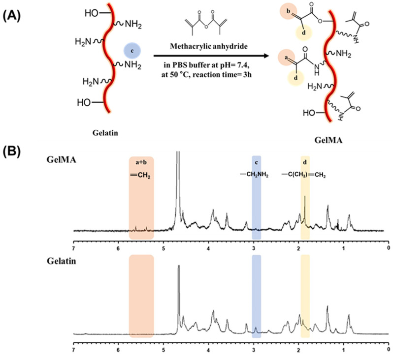
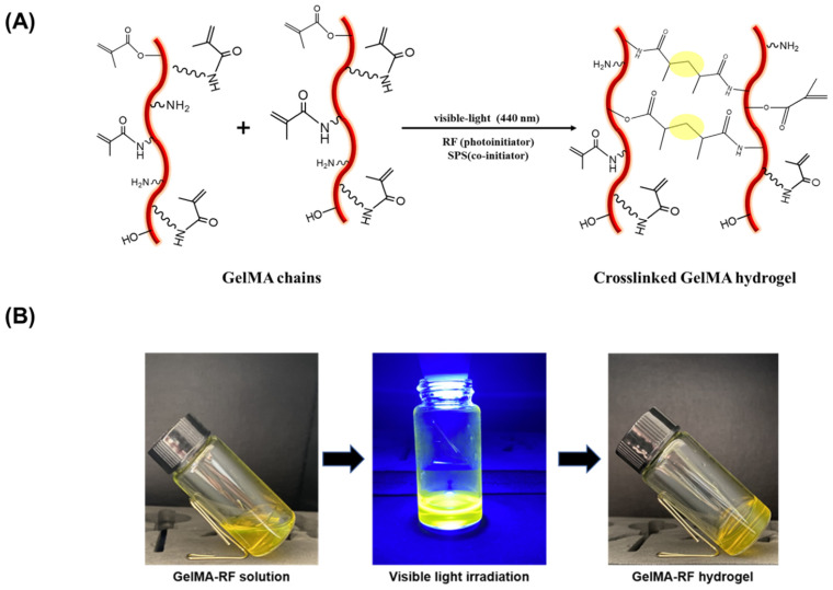
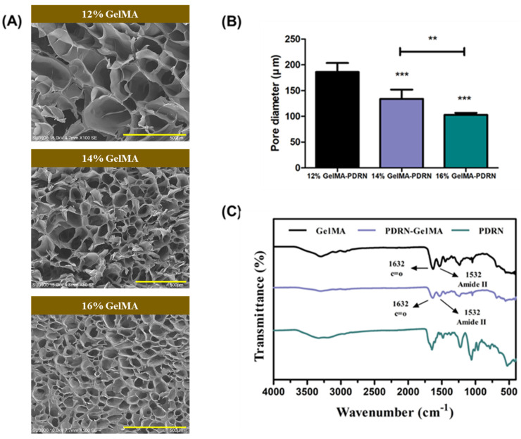
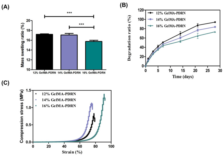
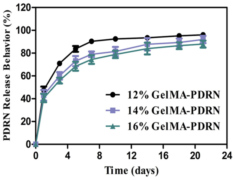
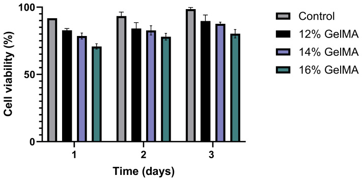
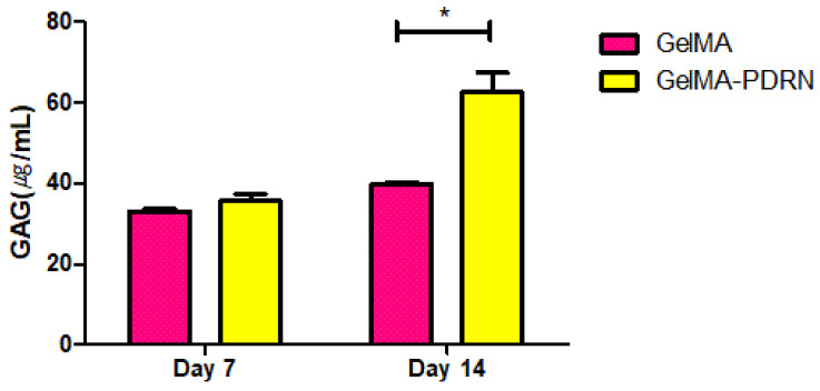
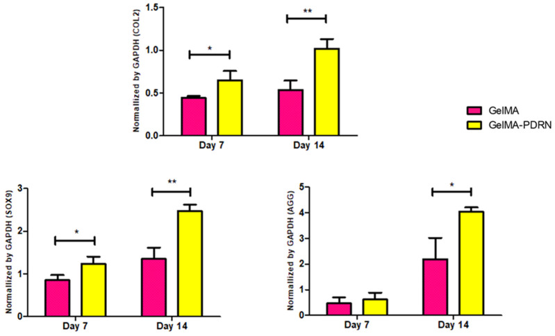
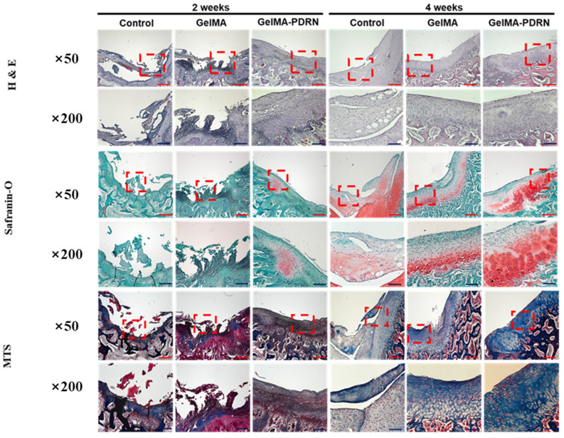
Articular cartilage faces challenges in self-repair due to the lack of blood vessels and limited chondrocyte concentration. Polydeoxyribonucleotide (PDRN) shows promise for promoting chondrocyte growth and cartilage regeneration, but its delivery has been limited to injections. Continuous PDRN delivery is crucial for effective cartilage regeneration. This study explores using gelatin methacrylate (gelMA) hydrogel, crosslinked with visible light and riboflavin 5'-phosphate sodium (RF) as a photoinitiator, for sustained PDRN release. GelMA hydrogel's synthesis was confirmed through spectrophotometric techniques, demonstrating successful methacrylate group incorporation. PDRN-loaded gelMA hydrogels displayed varying pore sizes, swelling ratios, degradation rates, and mechanical properties based on gelMA concentration. They showed sustained PDRN release and biocompatibility, with the 14% gelMA-PDRN composition performing best. Glycosaminoglycan (GAG) activity was higher in PDRN-loaded hydrogels, indicating a positive effect on cartilage formation. RT-PCR analysis revealed increased expression of cartilage-specific genes (COL2, SOX9, AGG) in gelMA-PDRN. Histological assessments in a rabbit cartilage defect model demonstrated superior regenerative effects of gelMA-PDRN hydrogels. This study highlights the potential of gelMA-PDRN hydrogels in cartilage tissue engineering, providing a promising approach for effective cartilage regeneration.

Keywords: GelMA hydrogel; PDRN; biocompatibility; cartilage regeneration; tissue engineering; visible light crosslinking.


Conflict of interest statement

The authors declare no conflict of interest.

Figures

Figure 1
Figure 1
Schematic representation shows (A) Synthesis of gelMA with addition of methacrylic anhydride to gelatin (B) 1H NMR spectra of gelatin and gelMA demonstrating successful functionalization.
Figure 2
Figure 2
(A) Schematic illustration of gelMA hydrogel photo-crosslinking reaction (B) Image of the formation of gelMA-RF hydrogel under visible light for 30 s.
Figure 3
Figure 3
(A) SEM images of cross-sections of gelMA hydrogel and gelMA-PDRN hydrogel, (B) The average pore size counted by ImageJ based on SEM images (scale bar = 500 μm) (mean ± SD, n = 3, ** p < 0.01, *** p < 0.001) (C) FT-IR spectra of PDRN only, gelMA, and gelMA-PDRN hydrogel.
Figure 4
Figure 4
Mechanical properties of gelMA-PDRN hydrogels (A) Mass swelling of gelMA-PDRN hydrogels (mean ± SD, n = 6) (B) Degradation ratio analyzed for 28 days (mean ± SD, n = 6) (C) Strain–stress curve of gelMA-PDRN hydrogel (mean ± SD, n = 3) (*** p < 0.001).
Figure 5
Figure 5
PDRN release profile of the gelMA-PDRN hydrogels with different gelMA concentrations (mean ± SD, n = 6).
Figure 6
Figure 6
Cell viability of examined GelMA-PDRN hydrogels by MTT assay for 3 days (mean ± SD, n = 6).
Figure 7
Figure 7
Biochemical characterization with GAG quantitative analysis (mean ± SD, n = 6, * p < 0.05).
Figure 8
Figure 8
Cartilage specific gene expression evaluated by RT-PCR with COL2, SOX9, and AGG normalized by GAPDH (mean ± SD, n = 6, * p < 0.05, ** p < 0.01).
Figure 9
Figure 9
Histology sections of all groups of the control, GelMA, and GelMA-PDRN groups were displayed at 2 and 4 weeks at 50× (scale bars = red 200 μm) and 200× (scale bars = blue 40 μm) magnifications with H&E, Safranin O, and MTS staining.

Similar articles

Cited by

References

    1. Huey D.J., Hu J.C., Athanasiou K.A. Unlike bone, cartilage regeneration remains elusive. Science. 2012;338:917–921. doi: 10.1126/science.1222454. - DOI - PMC - PubMed
    1. Kilmer C.E., Battistoni C.M., Cox A., Breur G.J., Panitch A., Liu J.C. Collagen type I and II blend hydrogel with autologous mesenchymal stem cells as a scaffold for articular cartilage defect repair. ACS Biomater. Sci. Eng. 2020;6:3464–3476. doi: 10.1021/acsbiomaterials.9b01939. - DOI - PMC - PubMed
    1. Krishnan Y., Grodzinsky A.J. Cartilage diseases. Matrix Biol. 2018;71:51–69. doi: 10.1016/j.matbio.2018.05.005. - DOI - PMC - PubMed
    1. Spahn G., Hofmann G.O., Klinger H.M. The effects of arthroscopic joint debridement in the knee osteoarthritis: Results of a meta-analysis. Knee Surg. Sports Traumatol. Arthrosc. 2013;21:1553–1561. doi: 10.1007/s00167-012-2169-1. - DOI - PubMed
    1. Riedl M., Vadalà G., Papalia R., Denaro V. Three-dimensional, scaffold-free, autologous chondrocyte transplantation: A systematic review. Orthop. J. Sports Med. 2020;8:2325967120951152. doi: 10.1177/2325967120951152. - DOI - PMC - PubMed

LinkOut - more resources