. 2025 Jan 6;15(1):22.
doi: 10.3390/bios15010022.

The Pre-Polarization and Concentration of Cells near Micro-Electrodes Using AC Electric Fields Enhances the Electrical Cell Lysis in a Sessile Drop

Affiliations

The Pre-Polarization and Concentration of Cells near Micro-Electrodes Using AC Electric Fields Enhances the Electrical Cell Lysis in a Sessile Drop

Kishor Kaphle et al. Biosensors (Basel). .

Abstract

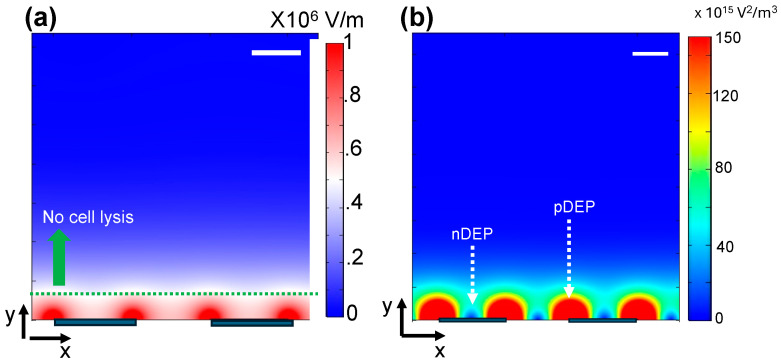
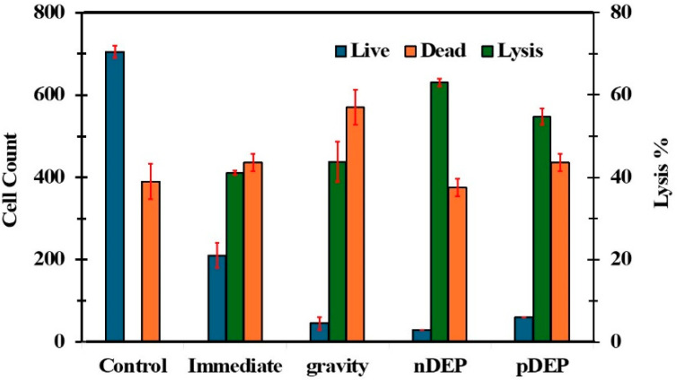
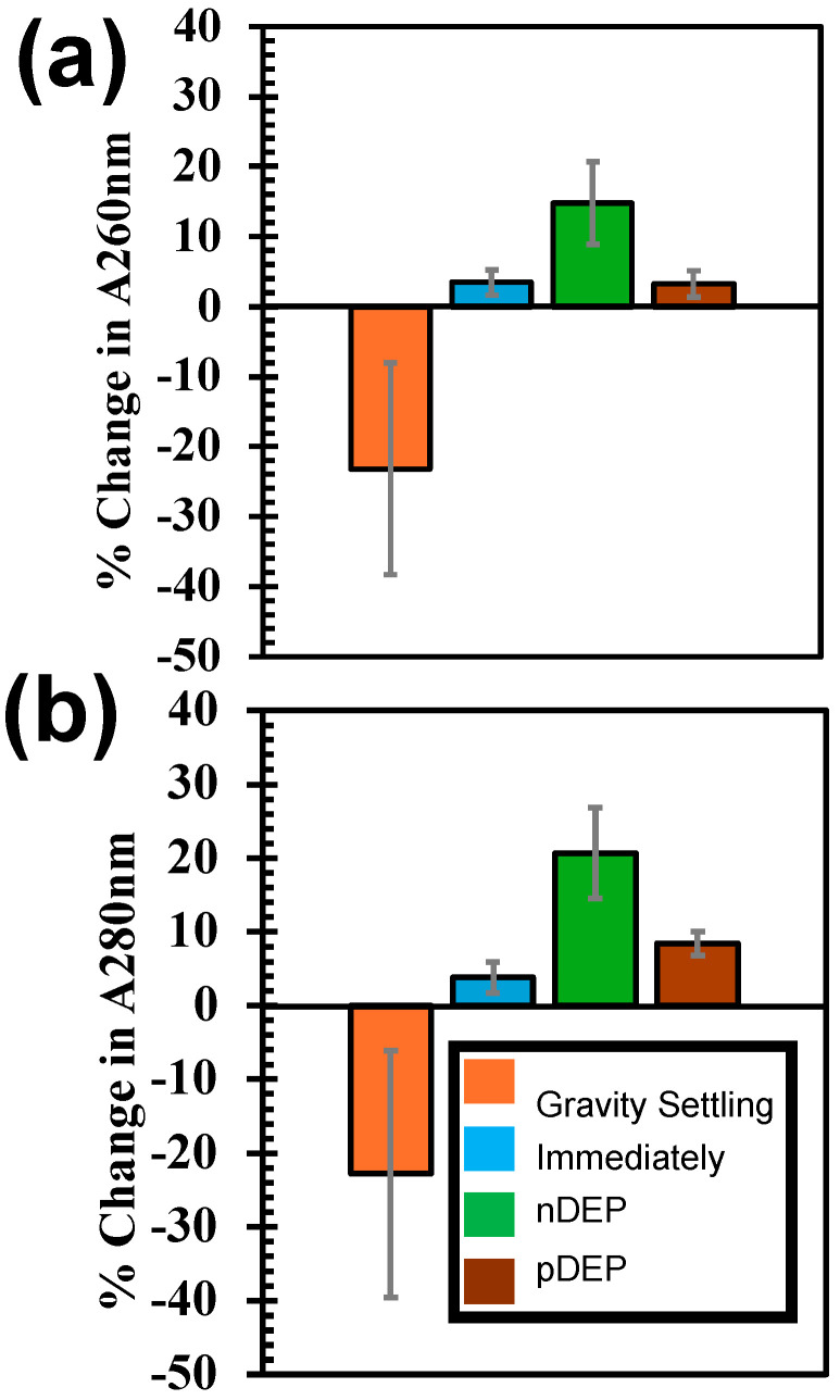
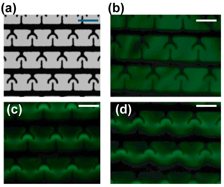
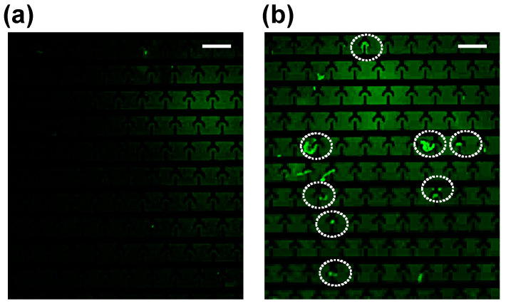
Cell lysis is the starting step of many biomedical assays. Electric field-based cell lysis is widely used in many applications, including point-of-care (POC) applications, because it provides an easy one-step solution. Many electric field-based lysis methods utilize micro-electrodes to apply short electric pulses across cells. Unfortunately, these cell lysis devices produce relatively low cell lysis efficiency as electric fields do not reach a significant portion of cells in the sample. Additionally, the utility of syringe pumps for flow cells in and out of the microfluidics channel causes cell loss and low throughput cell lysis. To address these critical issues, we suspended the cells in a sessile drop and concentrated on the electrodes. We used low-frequency AC electric fields (1 Vpp, 0-100 kHz) to drive the cells effectively towards electrodes and lysed using a short pulse of 10 V. A post-lysis analysis was performed using a hemocytometer, UV-vis spectroscopy, and fluorescence imaging. The results show that the pre-electric polarization of cells, prior to applying short electrical pulses, enhances the cell lysis efficiency. Additionally, the application of AC electric fields to concentrate cells on the electrodes reduces the assay time to about 4 min. In this study, we demonstrated that low-frequency AC electric fields can be used to pre-polarize and concentrate cells near micro-electrodes and improve cell lysis efficiency. Due to the simplicity and rapid cell lysis, this method may be suitable for POC assay development.

Keywords: cell lysis; dielectrophoresis and AC electroosmosis; electric fields; induced transmembrane potential.


Conflict of interest statement

The authors declare no conflicts of interest.

Figures

Figure 1
Figure 1
Calculated electric field (a) and electric field gradients (b) in the vicinity of the interdigitated electrodes used for cell lysis experiments. Scale bars indicate 5 µm.
Figure 2
Figure 2
Variation in cell lysis efficiency and cell count (after application lysis electric field) with experimental conditions used in the study. Control: cells from the tube left at room temperature; Immediate: immediately after pipetting cells on the electrodes; Gravity: settling cells under gravity; nDEP: after applying negative DEP and pDEP: after applying positive DEP.
Figure 3
Figure 3
The quantification of nucleic acid (a) and protein molecules (b) in the buffer after concentrating cells on the electrodes using gravity settling, immediately after pipetting cell sample on the electrodes, applying nDEP, pDEP. For all these conditions, the cell samples were lysed applying 10 V pulse for 2 s.
Figure 4
Figure 4
Concentration of cellular DNA on T-electrodes using AC electric fields. (a) Picture of interdigitated T-electrodes. (bd) fluorescence images of T-electrodes after applying no electric potential, 10 Vpp (1 MHz), 10 Vpp (500 kHz), respectively. Scale bars show 10 µm.
Figure 5
Figure 5
Concentration of cellular DNA on T-electrodes using AC electric fields (10 Vpp, 1 MHz). (a) Image of T-electrodes (from concentrated cells) without cell lysis. (b) Image of T-electrodes (from concentrated cells) on T-electrodes after electrical cell lysis. White circles show the concentrated DNA molecules on the electrodes. Scale bars show 20 µm.

Similar articles

References

    1. Morshed B.I., Shams M., Mussivand T. Electrical Lysis: Dynamics Revisited and Advances in On-Chip Operation. Crit. Rev. Biomed. Eng. 2013;41:37–50. doi: 10.1615/CritRevBiomedEng.2013006378. - DOI - PubMed
    1. Bahi M.M., Tsaloglou M.N., Mowlem M., Morgan H. Electroporation and lysis of marine microalga Karenia brevis for RNA extraction and amplification. J. R. Soc. Interface. 2011;8:601–608. doi: 10.1098/rsif.2010.0445. - DOI - PMC - PubMed
    1. Lu H., Schmidt M.A., Jensen K.F. A microfluidic electroporation device for cell lysis. Lab Chip. 2005;5:23–29. doi: 10.1039/b406205a. - DOI - PubMed
    1. Korohoda W., Grys M., Madeja Z. Reversible and irreversible electroporation of cell suspensions flowing through a localized DC electric field. Cell Mol. Biol. Lett. 2013;18:102–119. doi: 10.2478/s11658-012-0042-3. - DOI - PMC - PubMed
    1. Zhang Z., Zheng T., Zhu R. Single-cell individualized electroporation with real-time impedance monitoring using a microelectrode array chip. Microsyst. Nanoeng. 2020;6:81. doi: 10.1038/s41378-020-00196-0. - DOI - PMC - PubMed

LinkOut - more resources