. 2025 Jan 6;15(1):24.
doi: 10.3390/bios15010024.

A Truncated Multi-Thiol Aptamer-Based SARS-CoV-2 Electrochemical Biosensor: Towards Variant-Specific Point-of-Care Detection with Optimized Fabrication

Affiliations

A Truncated Multi-Thiol Aptamer-Based SARS-CoV-2 Electrochemical Biosensor: Towards Variant-Specific Point-of-Care Detection with Optimized Fabrication

Sergio Roberto Molina Ramirez et al. Biosensors (Basel). .

Abstract

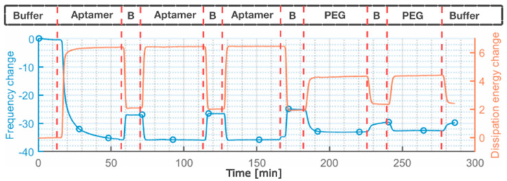
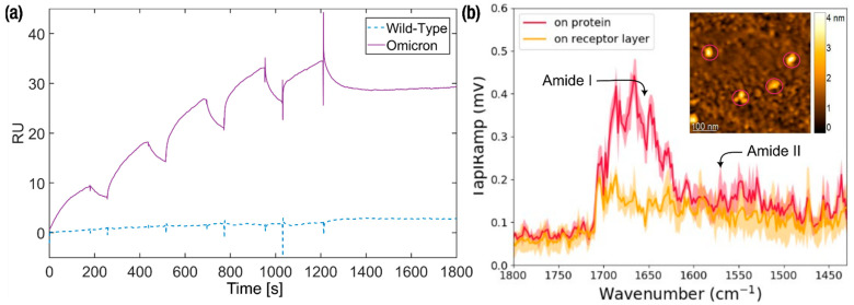
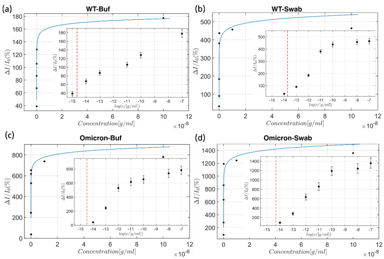
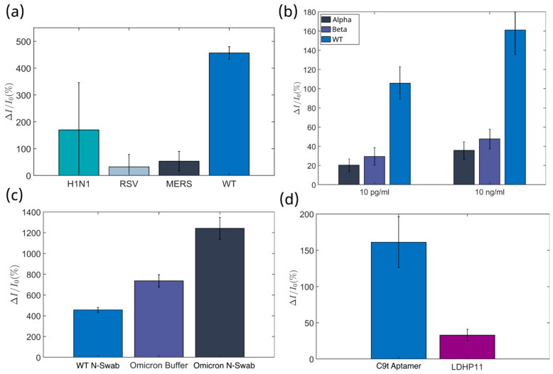
With the goal of fast and accurate diagnosis of infectious diseases, this study presents a novel electrochemical biosensor that employs a refined aptamer (C9t) for the detection of spike (S) protein SARS-CoV-2 variants in a flexible multielectrode aptasensor array with PoC capabilities. Two aptamer modifications were employed: removing the primer binding sites and including two dithiol phosphoramidite anchor molecules. Thus, reducing fabrication time from 24 to 3 h and increasing the stability and sparseness for multi-thiol aptasensors compared to a standard aptasensor using single thiols, without a reduction in aptamer density. The biosensor fabrication, optimization, and detection were verified in detail by electrochemistry, QCM-D, SPR, and XPS. The analyte-receptor binding was further confirmed spectroscopically at the level of individual molecules by AFM-IR. The aptasensor possesses a low limit of detection (8.0 fg/mL), the highest sensitivity reported for S protein (209.5 signal per concentration decade), and a wide dynamic detection range (8.0 fg/mL-38 ng/mL) in nasopharyngeal samples, covering the clinically relevant range. Furthermore, the C9t aptasensor showed high selectivity for SARS-CoV-2 S proteins over biomarkers for MERS-CoV, RSV, and Influenza. Even more, it showed a three times higher sensitivity for the Omicron in comparison to the Wuhan strain (wild type), alpha, and beta variants of the SARS-CoV-2 virus. Those results demonstrate the creation of an affordable and variant-selective refined C9t aptasensor that outperformed current rapid diagnosis tests.

Keywords: S protein; SARS-CoV-2; aptamer truncation; electrochemical aptasensor; multi-thiol.


Conflict of interest statement

The authors declare no conflicts of interest.

Figures

Figure 1
Figure 1
QCM-D measurements show the change of frequency in blue, and dissipation energy in orange. The aptamer solution flowed into the chamber in a 30 min window and was then cleaned with a buffer for 10 min. This procedure was repeated three times. For the PEG solution, two of these procedures were conducted. No change after the first buffer cleaning was observed for either. B stands for Buffer.
Figure 2
Figure 2
SPR and AFM-IR investigations. (a) SPR measurements with concentration series of 55, 166, 500, 1500, and 3000 nM of the truncated C9t aptamer for binding affinity determination to wild-type (dash line) and Omicron (continuous line) S proteins. (b) AFM-IR topography (inset) and spectra of aptamer C9t/PEG layer and S protein. The inset shows the AFM height image with the S protein–C9t aptamer complexes (circled) resolved. Spectra recorded of the S protein–C9t complex (circled) exhibit a strong feature assigned to the amide I band, followed by a broad feature at lower wave numbers assigned to the amide II band of protein. The spectra are averaged from the four circled positions and four positions on the receptor layer.
Figure 3
Figure 3
Calibration curves. The calibration curves represent the response of the aptasensor in a concentration ranging from 1 fg/mL to 100 ng/mL of the target proteins: against (a) wild-type S protein of the SARS-CoV-2 virus in ferri/ferrocyanide solution and (b) wild-type S protein of the SARS-CoV-2 in a spiked negative control (non-infectious) nasal swab, (c) the S protein of the Omicron variant in ferri/ferrocyanide solution, and (d) the S protein of the Omicron variant in a spiked negative (non-infectious) control nasal swab. The overlay figure shows the Langmuir–Freundlich curve representation of the biosensor’s response without logarithmic axis modifications, whilst the inset shows the logarithmic representation of the obtained data, and the dashed line represents the LoD for the individual media–analyte combination. Hereby, all calibration curves were determined at 16 individual electrodes per chip, for three chips per condition.
Figure 4
Figure 4
Selectivity Tests for the Aptasensor. The aptasensor’s response to analog analytes (selectivity tests) was conducted at an analyte concentration of 10 ng/mL. The selectivity test was performed as the responses to (a) wild-type response for viral proteins of other respiratory viruses with similar symptoms, (b) different strains of the SARS-CoV-2 virus, and (c) between wild-type and the Omicron variant, in ferri/ferrocyanide solution (Buffer), as well as in spiked negative nasal swab samples (N-Swab). (d) Comparison of the detection response in buffer for Omicron S protein by the C9t aptasensor versus a previously reported malaria aptamer LDHp11 [54]. Hereby, all selectivity tests were conducted at 16 individual electrodes per chip, for two chips per condition.

Similar articles

Cited by

References

    1. Morens D.M., Breman J.G., Calisher C.H., Doherty P.C., Hahn B.H., Keusch G.T., Kramer L.D., LeDuc J.W., Monath T.P., Taubenberger J.K. The Origin of COVID-19 and Why It Matters. Am. J. Trop. Med. Hyg. 2020;103:955–959. doi: 10.4269/ajtmh.20-0849. - DOI - PMC - PubMed
    1. Morens D.M., Fauci A.S. Emerging Pandemic Diseases: How We Got to COVID-19. Cell. 2020;182:1077–1092. doi: 10.1016/j.cell.2020.08.021. - DOI - PMC - PubMed
    1. Cui J., Li F., Shi Z.-L. Origin and evolution of pathogenic coronaviruses. Nat. Rev. Microbiol. 2019;17:181–192. doi: 10.1038/s41579-018-0118-9. - DOI - PMC - PubMed
    1. Souf S. Recent Advances in diagnostic testing for viral infections. Biosci. Horiz. 2016;9:hzw010. doi: 10.1093/biohorizons/hzw010. - DOI
    1. Aylward B., Liang W., Dong X., Eckmanns T., Fisher D., Ihekweazu C., Lane C., Lee J.-K., Leung G., Lin J., et al. WHO China Joint Mission on COVID-19 Final Report. 2020. [(accessed on 21 November 2024)]. Available online: https://www.who.int/docs/default-source/coronaviruse/who-china-joint-mis....

MeSH terms

Substances

Supplementary concepts

LinkOut - more resources