Review
. 2024 Dec 31;12(1):14.
doi: 10.3390/jcdd12010014.

Papillary Muscles of the Left Ventricle: Integrating Electrical and Mechanical Dynamics

Affiliations
Review

Papillary Muscles of the Left Ventricle: Integrating Electrical and Mechanical Dynamics

Csilla Andrea Eötvös et al. J Cardiovasc Dev Dis. .

Abstract

Background: Papillary muscles are structures integrated into the mitral valve apparatus, having both electrical and mechanical roles. The importance of the papillary muscles (PM) is mainly related to cardiac arrhythmias and mitral regurgitation. The aim of this review is to offer an overview of the anatomy and physiology of the papillary muscles, along with their involvement in cardiovascular pathologies, including arrhythmia development in various conditions and their contribution to secondary mitral regurgitation.

Methods: A literature search was performed on PubMed using the following relevant keywords: papillary muscles, mitral valve, arrhythmia, anatomy, and physiology.

Results: During the cardiac cycle, papillary muscles have continuous dimensional and pressure changes. On one hand, their synchrony or dyssynchrony impacts the process of mitral valve opening and closure, and on the other hand, the pressure changes can trigger electrical instability. There is increased awareness of papillary muscles as an arrhythmic source. Arrhythmias arising from PM were found in patients with or without structural heart disease, via Purkinje fibres, due to increased automaticity or triggered activity.

Conclusions: Despite the interest in mitral valve physiology, there are still many unknowns in relation to the papillary muscles, especially with regard to their role in arrhythmogenesis and the pathogenesis of mitral regurgitation.

Keywords: arrhythmia; dyssynchrony; mitral regurgitation; mitral valve; papillary muscles.


Conflict of interest statement

The authors declare no conflicts of interest.

Figures
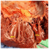
Figure 1
Figure 1
Anterolateral (APM) and posteromedial (PPM) papillary muscles in a necropsy specimen (courtesy of Dr. Doinita Crisan).
Figure 2
Figure 2
Papillary muscle (PM) length variation during cardiac cycle in contrast with left ventricular wall (LV wall). The electrical activation of the left ventricular papillary muscles occurs during the early stages of ventricular activation, while their mechanical contraction happens about 20 ms after the contraction of the surrounding epicardium (Armour et al. [17]).
Figure 3
Figure 3
Characteristic electrocardiographic pattern for ectopy originating from the posteromedial papillary muscle. The superior axis and qR pattern, with slurred R wave and short intrinsic deflection (66 ms), in lead V1 is notable. In addition, there is a late transition in the precordial leads. Electrocardiogram from a patient with mitral valve prolapse.
Figure 4
Figure 4
Elements involved in genesis of mitral regurgitation: atrial dilation, annulus dilation, papillary muscle dyssynchrony, left ventricular dilation. (LA: left atrium, MV: mitral valve, Ant: anterior mitral leaflet, Post: posterior mitral leaflet, LVOT: left ventricular outflow tract, PM: posterior papillary muscle, AL: anterior papillary muscle).
Figure 5
Figure 5
(A) PPM in parasternal long axis view (yellow arrow) in a patient with mitral valve prolapse. (B) Parasternal mid-ventricular short axis view at the level of PMs. Notice that PPM has two heads (red arrow), while APM has one (white arrow). (PPM: postero-medial papillary muscle; APM: antero-lateral papillary muscle, PM: papillary muscle).
Figure 6
Figure 6
Cardiac MRI. PM hypertrophy in a patient with HOCM (yellow arrows). (MRI: magnetic resonance imaging, HOCM: hypertrophic obstructive cardiomyopathy, PM: papillary muscle).

References

    1. Clayton M.P.R. Leonardo da Vinci: Anatomist. 2012nd ed. Volume 1. Royal Collection Trust; London, UK: 2014. p. 260.
    1. Madu E.C., D’Cruz I.A. The vital role of papillary muscles in mitral and ventricular function: Echocardiographic insights. Clin. Cardiol. 1997;20:93–98. doi: 10.1002/clc.4960200203. - DOI - PMC - PubMed
    1. Naksuk N., Kapa S., Asirvatham S.J. Spectrum of Ventricular Arrhythmias Arising from Papillary Muscle in the Structurally Normal Heart. Card. Electrophysiol. Clin. 2016;8:555–565. doi: 10.1016/j.ccep.2016.04.005. - DOI - PubMed
    1. Roberts W.C., Cohen L.S. Left ventricular papillary muscles. Description of the normal and a survey of conditions causing them to be abnormal. Circulation. 1972;46:138–154. doi: 10.1161/01.CIR.46.1.138. - DOI - PubMed
    1. Saha A., Roy S. Papillary muscles of left ventricle-Morphological variations and it’s clinical relevance. Indian Heart J. 2018;70:894–900. doi: 10.1016/j.ihj.2017.12.003. - DOI - PMC - PubMed

LinkOut - more resources