Dermoscopy and Ultraviolet-Enhanced Fluorescence Dermoscopy (UEFD) Increase the Accuracy of Diagnosis and Are Useful in Assessing the Effectiveness of Kerion celsi Treatment
- PMID: 39852471
- PMCID: PMC11766397
- DOI: 10.3390/jof11010052
Dermoscopy and Ultraviolet-Enhanced Fluorescence Dermoscopy (UEFD) Increase the Accuracy of Diagnosis and Are Useful in Assessing the Effectiveness of Kerion celsi Treatment
Abstract
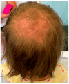
Microsporum canis, a zoophilic dermatophyte, infects the stratum corneum and keratinized tissues like hair and nails in cats and dogs, with cats serving as the primary reservoir. Most human infections arise from animal contact. We present the case of a girl aged 8 with skin scalp lesions persisting for two months. Several scalp lesions, with a maximum diameter of 4 cm, presented as erythematous plaques with superficial scaling, yellow crusts, and edematous areas with purulent exudate. Dermoscopy revealed yellow crusts on an erythematous background, along with white scales, pustules, broken hairs, and comma hairs. Ultraviolent-enhanced fluorescence dermoscopy (UEFD) showed slight celadon green fluorescence, which enhanced the diagnosis and further helped to monitor the treatment. The PCR test confirmed the presence of M. canis. Treatment included topical ciclopirox and oral terbinafine. Lesions on the scalp and noticeable hair regrowth were observed in the areas of hair loss after two months. Kerion celsi can result in severe alopecia. To prevent scarring associated with hair loss in children, early mycological diagnostics, supported by dermoscopy and UEFD, is recommended.
Keywords: Kerion celsi; Microsporum canis; alopecia; dermatophyte; fungal infection; zoonosis.
Conflict of interest statement
The authors declare no conflicts of interest.
Figures


References
Publication types
LinkOut - more resources
Full Text Sources
Miscellaneous

