. 2025 Jan 14;11(1):58.
doi: 10.3390/jof11010058.

Direct and Indirect Protein Interactions Link FUS Aggregation to Histone Post-Translational Modification Dysregulation and Growth Suppression in an ALS/FTD Yeast Model

Affiliations

Direct and Indirect Protein Interactions Link FUS Aggregation to Histone Post-Translational Modification Dysregulation and Growth Suppression in an ALS/FTD Yeast Model

Seth A Bennett et al. J Fungi (Basel). .

Abstract

Amyotrophic lateral sclerosis (ALS) and frontotemporal dementia (FTD) are incurable neurodegenerative disorders sharing pathological and genetic features, including mutations in the FUS gene. FUS is an RNA-binding protein that mislocalizes to the cytoplasm and aggregates in ALS/FTD. In a yeast model, FUS proteinopathy is connected to changes in the epigenome, including reductions in the levels of H3S10ph, H3K14ac, and H3K56ac. Exploiting the same model, we reveal novel connections between FUS aggregation and epigenetic dysregulation. We show that the histone-modifying enzymes Ipl1 and Rtt109-responsible for installing H3S10ph and H3K56ac-are excluded from the nucleus in the context of FUS proteinopathy. Furthermore, we found that Ipl1 colocalizes with FUS, but does not bind it directly. We identified Nop1 and Rrp5, a histone methyltransferase and rRNA biogenesis protein, respectively, as FUS binding partners involved in the growth suppression phenotype connected to FUS proteinopathy. We propose that the nuclear exclusion of Ipl1 through indirect interaction with FUS drives the dysregulation of H3S10ph as well as H3K14ac via crosstalk. We found that the knockdown of Nop1 interferes with these processes. In a parallel mechanism, Rtt109 mislocalization results in reduced levels of H3K56ac. Our results highlight the contribution of epigenetic mechanisms to ALS/FTD and identify novel targets for possible therapeutic intervention.

Keywords: FUS; Ipl1; Nop1; Rtt109; amyotrophic lateral sclerosis; epigenetics; frontotemporal dementia; histone post-translational modifications.


Conflict of interest statement

The authors declare no conflicts of interest.

Figures
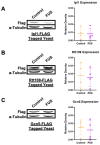
Figure 1
Figure 1
Levels of select histone-modifying enzymes remain unchanged in connection to FUS proteinopathy. The levels of (A) Ipl1, (B) Rtt109, and (C) Gcn5 were measured through immunoblotting against FLAG in yeast overexpressing a control (orange) or FUS (purple) vector. α-Tubulin was used as a loading control. Column scatterplots compiling multiple independent biological replicates display the mean fold change in the FLAG expression based on densitometric analysis. Error bars represent ±SD. n = 4.
Figure 2
Figure 2
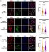
Ipl1 and Rtt109 are depleted from the nucleus in FUS proteinopathy yeast models. FUS or control yeast expressing (A) Ipl1-FLAG (n = 368 controls, 360 FUS), (B) Rtt109-FLAG (n = 199 controls, 235 FUS), or (C) Gcn5-FLAG (n = 348 controls, 315 FUS) were imaged using immunofluorescence with antibodies recognizing FLAG (red) and FUS (green) and counterstained with DAPI (blue). Column scatterplots represent the percent of the FLAG signal in the nucleus. Examples of Ipl1-FLAG and FUS colocalization are highlighted with white arrows. **** = p < 0.0001.
Figure 3
Figure 3
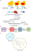
Putative binding partners of FUS are involved in rRNA processing and ATP binding. (A) Schematic representation of co-immunoprecipitation experiments using an FUS antibody as bait. Negative controls are also shown. (B) Diagram portraying filtering of FUS Co-IP protein hits. (C) Enrichment map created from GO annotations and KEGG Pathways associated with putative FUS yeast binding partners. Nodes highlighted in the yellow oval correspond to annotations related to ATP binding, and nodes highlighted in the teal circle correspond to annotations involved in rRNA processing.
Figure 4
Figure 4
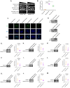
Reduced levels of either Rrp5 or Nop1 mRNA relieve growth suppression but show differential effects on histone PTM levels in FUS-overexpressing yeast. (A) Serial growth dilution assays depicted cell viability of parental, Rrp5 DAmP, and Nop1 DAmP control and FUS overexpression lines spotted on glucose (FUS “off”) or galactose (FUS “on”) media (n = 3). (B) Column scatterplot represents densitometric measurement of cell density of FUS yeast (middle spot) compared to control yeast on galactose plates in (A). *** = p < 0.001; **** = p < 0.0001. (C) Parental, Rrp5 DAmP, and Nop1 DAmP FUS or control yeast were imaged using immunofluorescence with antibodies against FUS (green) and counterstained with DAPI (blue). (D) Western blots confirmed the expression of FUS in these cells. n = 3. The levels of (E) H3S10ph, (F) H3K14ac, and (G) H3K56ac were measured in control (orange) and FUS (purple) parental yeast through immunoblotting. Similarly, levels of (H) H3S10ph, (I) H3K14ac, and (J) H3K56ac were measured in Rrp5 DAmP control and FUS yeast. Finally, levels of (K) H3S10ph, (L) H3K14ac, and (M) H3K56ac were measured in Nop1 DAmP control and FUS yeast. Column scatterplots compiling multiple biological replicates display the densities of histone post-translational modifications relative to the density of histone H3 as a loading control. Error bars represent ±SD. n = 4. * = p < 0.05; *** = p < 0.001.
Figure 5
Figure 5
Reduced Rrp5 or Nop1 mRNA levels do not affect TDP-43 overexpression levels, growth suppression, or histone PTMs. (A) Serial growth dilution assays depicted cell viability of parental, Rrp5 DAmP, and Nop1 DAmP control and TDP-43 overexpression lines spotted on glucose (TDP-43 “off”) or galactose (TDP-43 “on”) media (n = 3). (B) Column scatterplot represents densitometric measurement of cell density of TDP-43 yeast (middle spot) compared to control yeast on galactose plates in (A). (C) Western blots confirm the expression of TDP-43 in parental cells. The levels of (D) H3S10ph, (E) H3K14ac, and (F) H3K56ac were measured in control (orange) and TDP-43 (purple) parental yeast through immunoblotting. Similarly, (G) expression of TDP-43 as well as levels of (H) H3S10ph, (I) H3K14ac, and (J) H3K56ac were measured in Rrp5 DAmP control and TDP-43 yeast. Finally, (K) expression of TDP-43 and levels of (L) H3S10ph, (M) H3K14ac, and (N) H3K56ac were measured in Nop1 DAmP control and TDP-43 yeast. Column scatterplots compiling multiple biological replicates display the densities of histone post-translational modifications relative to the density of histone H3 as a loading control. Error bars represent ±SD. n = 3–7.
Figure 6
Figure 6
Putative mechanisms linking histone PTMs to FUS proteinopathy in yeast. Ipl1 is excluded from the nucleus through an indirect interaction with FUS, leading to reduced levels of H3S10ph. The H3K14ac levels are likely lowered through histone crosstalk with H3S10ph. A direct interaction between FUS and either Rrp5 or Nop1 is linked to cytotoxicity, while FUS’s interaction with Nop1 connects to changes in H3S10ph and H3K14ac. In a parallel mechanism, Rtt109 mislocalization contributes to the decrease in H3K56ac levels. All these associations do not occur in the context of TDP-43 proteinopathy and hence are not related to protein aggregation in general.

Similar articles

Cited by

References

    1. Pang S.Y.-Y., Hsu J.S., Teo K.-C., Li Y., Kung M.H.W., Cheah K.S.E., Chan D., Cheung K.M.C., Li M., Sham P.-C., et al. Burden of Rare Variants in ALS Genes Influences Survival in Familial and Sporadic ALS. Neurobiol. Aging. 2017;58:238.e9–238.e15. doi: 10.1016/j.neurobiolaging.2017.06.007. - DOI - PubMed
    1. Nolan M., Talbot K., Ansorge O. Pathogenesis of FUS-Associated ALS and FTD: Insights from Rodent Models. Acta Neuropathol. Commun. 2016;4:99. doi: 10.1186/s40478-016-0358-8. - DOI - PMC - PubMed
    1. Ferrari R., Kapogiannis D., Huey E.D., Momeni P. FTD and ALS: A Tale of Two Diseases. Curr. Alzheimer Res. 2011;8:273–294. doi: 10.2174/156720511795563700. - DOI - PMC - PubMed
    1. Debove C., Zeisser P., Salzman P.M., Powe L.K., Truffinet P. The Rilutek (Riluzole) Global Early Access Programme: An Open-Label Safety Evaluation in the Treatment of Amyotrophic Lateral Sclerosis. Amyotroph. Lateral Scler. Other Mot. Neuron Disord. 2001;2:153–158. doi: 10.1080/146608201753275508. - DOI - PubMed
    1. Brooks B.R., Jorgenson J.A., Newhouse B.J., Shefner J.M., Agnese W. Edaravone in the Treatment of Amyotrophic Lateral Sclerosis: Efficacy and Access to Therapy—A Roundtable Discussion. Am. J. Manag. Care. 2018;24:S175–S186. - PubMed

LinkOut - more resources