. 2025 Jan 13;13(1):70.
doi: 10.3390/vaccines13010070.

T4 Phage Displaying Dual Antigen Clusters Against H3N2 Influenza Virus Infection

Affiliations

T4 Phage Displaying Dual Antigen Clusters Against H3N2 Influenza Virus Infection

Shenglong Liu et al. Vaccines (Basel). .

Abstract

Background: The current H3N2 influenza subunit vaccine exhibits weak immunogenicity, which limits its effectiveness in preventing and controlling influenza virus infections.

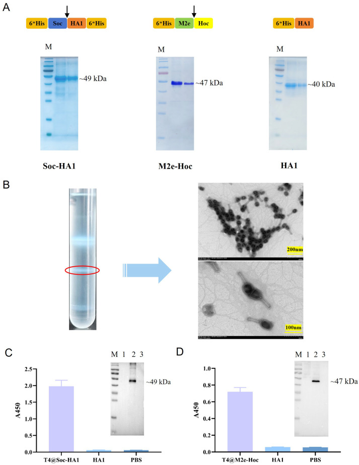
Methods: In this study, we aimed to develop a T4 phage-based nanovaccine designed to enhance the immunogenicity of two antigens by displaying the HA1 and M2e antigens of the H3N2 influenza virus on each phage nanoparticle. Specifically, we fused the Soc protein with the HA1 antigen and the Hoc protein with the M2e antigen, assembling them onto a T4 phage that lacks Soc and Hoc proteins (Soc-Hoc-T4), thereby constructing a nanovaccine that concurrently presents both HA1 and M2e antigens.

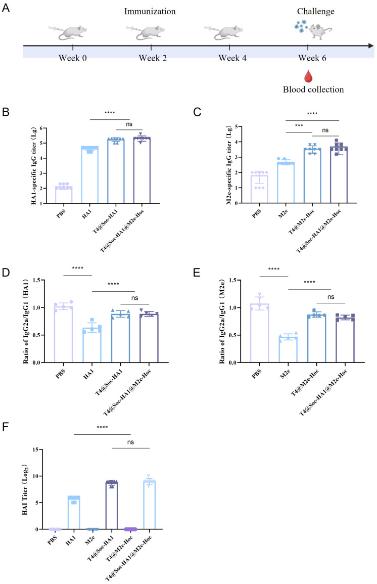
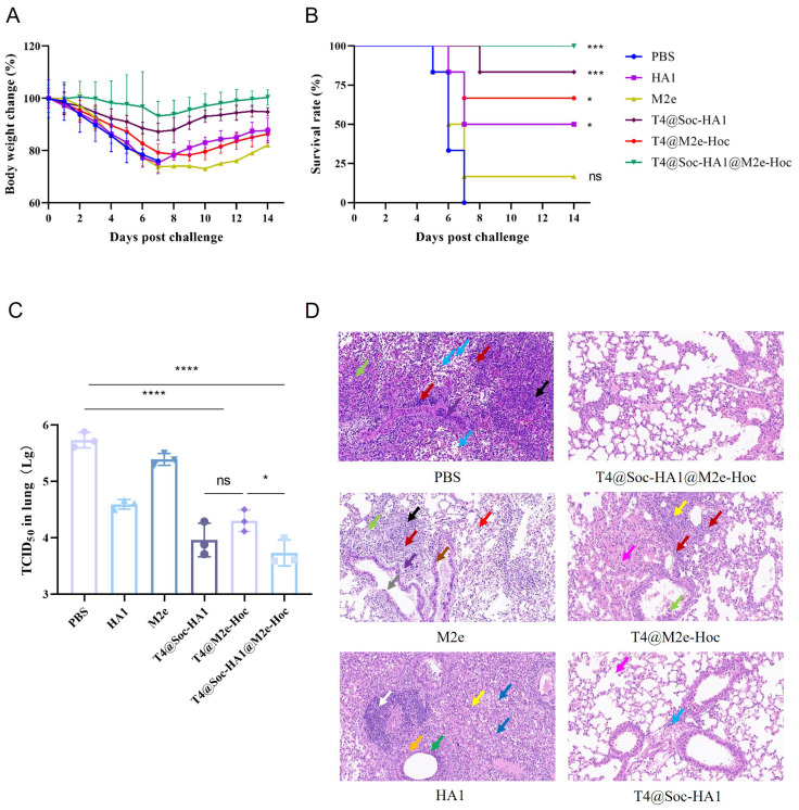
Results: The analysis of the optical density of the target protein bands indicated that each particle could display approximately 179 HA1 and 68 M2e antigen molecules. Additionally, animal experiments demonstrated that this nanoparticle vaccine displaying dual antigen clusters induced a stronger specific immune response, higher antibody titers, a more balanced Th1/Th2 immune response, and enhanced CD4+ and CD8+ T cell effects compared to immunization with HA1 and M2e antigen molecules alone. Importantly, mice immunized with the T4 phage displaying dual antigen clusters achieved full protection (100% protection) against the H3N2 influenza virus, highlighting its robust protective efficacy.

Conclusions: In summary, our findings indicate that particles based on a T4 phage displaying antigen clusters exhibit ideal immunogenicity and protective effects, providing a promising strategy for the development of subunit vaccines against various viruses beyond influenza.

Keywords: HA1 and M2e antigen molecule clusters; T4 phage nanovaccine; immune response.


Conflict of interest statement

The authors have declared that no competing interests exist.

Figures

Scheme 1
Scheme 1
Schematic diagram of T4 phage-based nanovaccine displaying dual antigen clusters. The HA1 and M2e antigens of the H3N2 influenza virus were fused with the Soc and Hoc protein to generate the Soc-HA1 and M2e-Hoc fusion proteins, respectively. These two fusion proteins were co-incubated in vitro with the SocHocT4, resulting in the formation of the T4@Soc-HA1@M2e-Hoc nanovaccine.
Figure 1
Figure 1
Construction of the nanovaccine. (A) Expression and purification of Soc-HA1, M2e-Hoc, and HA1 proteins. The black arrows indicate flexible linkers between the two proteins (M: marker). (B) Purification and TEM characterization of SocHocT4 phage. The red circle denotes the purified SocHocT4 phage, while the arrow indicates the characterization of this SocHocT4 phage under TEM. (C) Validation of in vitro assembly of T4@Soc-HA1 nanoparticles; confirmed using ELISA and Western blot analysis (Lane M: marker, Lane 1: SocHocT4 phage, Lane 2: T4@Soc-HA1 particles assembled and centrifuged pellet, Lane 3: supernatant after washing and centrifugation). (D) Validation of in vitro assembly of T4@M2e-Hoc nanoparticles; confirmed using ELISA and Western blot analysis (Lane M: marker, Lane 1: SocHocT4 phage, Lane 2: T4@M2e-Hoc particles assembled and centrifuged pellet, Lane 3: supernatant after washing and centrifugation).
Figure 2
Figure 2
HA1/M2e-specific humoral immune responses. (A) Immunization regimen. (B) HA1-specific IgG titers measured by ELISA. (C) M2e-specific IgG titers measured by ELISA. (D) Ratio of HA1-specific IgG2a to IgG1 in serum. (E) Ratio of M2e-specific IgG2a to IgG1 in serum. (F) HAI antibody titers in serum. Data are represented as mean ± S.D. ***, **** and ns indicate p < 0.001, p < 0.0001 and not statistically significant, respectively (ANOVA).
Figure 3
Figure 3
HA1/M2e-specific cellular immune responses. Mice were immunized according to the regimen shown in Figure 2A, and splenocytes were isolated on day 14 after the final immunization. (A) Splenocytes isolated from immunized mice were stained with anti-CD3, CD4, and CD8a antibodies and analyzed by flow cytometry. (B) Percentage of CD3+CD4+ T cells within the splenocyte population. (C) Percentage of CD3+CD8+ T cells within the splenocyte population. (D) Levels of IL-4 in the culture supernatant of splenocytes. (E) Levels of IFN-γ in the culture supernatant of splenocytes. Data are represented as mean ± S.D. **, ***, **** and ns indicate p < 0.01, p < 0.001, p < 0.0001 and not statistically significant, respectively (ANOVA).
Figure 4
Figure 4
Evaluation of nanovaccine protection efficacy. Mice (n = 6) were subjected to a viral challenge two weeks after the final immunization. (A) Changes in mice body weight over 14 days post-challenge. (B) Survival rates of mice over 14 days post-challenge. (C) Lung viral loads 5 days post-challenge. (D) Histopathological analysis of the lungs from virus-challenged mice (n = 3), which were immunized using the same regimen as described above. Mice were euthanized, and lung sections were prepared as described in the Materials and Methods. Images represent typical results for each group, including multifocal necrosis in lung tissue (black arrows), numerous fibroblasts (light green arrows), a heavy infiltration of lymphocytes and granulocytes (dark red arrows), slight granulocyte infiltration in the alveolar walls (red arrows), abundant lymphocytes, granulocytes, and macrophages within the alveoli (light blue arrows), slight bronchiolar epithelial cell necrosis (gray arrows), necrotic cell debris and eosinophilic material (brown arrows), slight perivascular edema (blue arrows), leukocytes occluding the lumen (purple arrows), necrotic cell debris (yellow arrows), detached epithelial cells (green arrows), a small amount of perivascular lymphocytic infiltration is observed in a ring-like pattern (white arrows), and mild hemorrhage (light purple arrows). The data were analyzed using the Mantel–Cox test. *, ***, **** and ns indicate p < 0.05, p < 0.001, p < 0.0001 and not statistically significant, respectively (ANOVA).

Similar articles

References

    1. Du X., King A.A., Woods R.J., Pascual M. Evolution-informed forecasting of seasonal influenza A (H3N2) Sci. Transl. Med. 2017;9:eaan5325. doi: 10.1126/scitranslmed.aan5325. - DOI - PMC - PubMed
    1. Taubenberger J.K., Morens D.M. The pathology of influenza virus infections. Annu. Rev. Pathol. 2008;3:499–522. doi: 10.1146/annurev.pathmechdis.3.121806.154316. - DOI - PMC - PubMed
    1. Smith G.J., Vijaykrishna D., Bahl J., Lycett S.J., Worobey M., Pybus O.G., Ma S.K., Cheung C.L., Raghwani J., Bhatt S., et al. Origins and evolutionary genomics of the 2009 swine-origin H1N1 influenza A epidemic. Nature. 2009;459:1122–1125. doi: 10.1038/nature08182. - DOI - PubMed
    1. Ferdinands J.M., Gaglani M., Martin E.T., Middleton D., Monto A.S., Murthy K., Silveira F.P., Talbot H.K., Zimmerman R., Alyanak E., et al. Prevention of Influenza Hospitalization Among Adults in the United States, 2015-2016: Results From the US Hospitalized Adult Influenza Vaccine Effectiveness Network (HAIVEN) J. Infect. Dis. 2019;220:1265–1275. doi: 10.1093/infdis/jiy723. - DOI - PMC - PubMed
    1. Muennig P.A., Khan K. Cost-effectiveness of vaccination versus treatment of influenza in healthy adolescents and adults. Clin. Infect. Dis. 2001;33:1879–1885. doi: 10.1086/324491. - DOI - PubMed

LinkOut - more resources