. 2024 Dec 29;12(1):8.
doi: 10.3390/vetsci12010008.

The Development of a One-Step PCR Assay for Rapid Detection of an Attenuated Vaccine Strain of Duck Hepatitis Virus Type 3 in Korea

Affiliations

The Development of a One-Step PCR Assay for Rapid Detection of an Attenuated Vaccine Strain of Duck Hepatitis Virus Type 3 in Korea

Cheng-Dong Yu et al. Vet Sci. .

Abstract

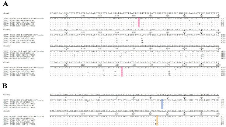
Duck hepatitis A virus type 3 (DHAV-3) is a viral pathogen that causes acute, high-mortality hepatitis in ducklings, and vaccination with attenuated live vaccines is currently the main preventive measure against it. However, differentiating infected from vaccinated animals (DIVA) is crucial for clinical diagnosis and effective disease control. This study aimed to develop a rapid mismatch amplification mutation assay PCR (MAMA-PCR) diagnostic method to simultaneously detect and differentiate between wild-type and vaccine strains. The method was specifically designed to target the critical single-nucleotide polymorphism (SNP) site (T→C at position 1143 in the VP0 gene) unique to the Korean vaccine strain AP04203-P100. MAMA-PCR demonstrated high sensitivity and specificity, with detection limits as low as 102.4 ELD50/mL for wild strains and 100.5 ELD50/mL for vaccine strains, and showed no cross-reactivity with 11 other common duck pathogens. The clinical sample results were completely consistent with those obtained using nested PCR detection and gold-standard sequencing. In summary, we successfully developed a rapid, one-step MAMA-PCR method that is more suitable for clinical diagnosis than traditional sequencing methods.

Keywords: differentiating infected from vaccinated animals; duck hepatitis A virus type 3; mismatch amplification mutation assay; rapid diagnostic.


Conflict of interest statement

Authors Hyung-Kwan Jang and Min Kang were employed by Bio Disease Control (BIOD) Co., Ltd. The remaining authors declare that the research was conducted in the absence of any commercial or financial relationships that could be construed as potential conflicts of interest.

Figures

Figure 1
Figure 1
Comparison of the VP0 gene between the Korean DHAV-3 vaccine strain AP04203-P100 and wild-type strains. The nucleotide and corresponding amino acid sequences of the vaccine strain and four known Korean wild-type strains from NCBI were aligned and analyzed using the MegAlign program (DNAStar Lasergene, version 11). (A) Two distinguishing SNP sites were identified at positions 853 and 1143 in the VP0 gene (highlighted in red). (B) The SNP at position 853 does not alter the amino acid, with both vaccine and wild-type strains retaining serine (Ser, S), indicating a synonymous mutation (highlighted in blue). In contrast, the SNP at position 1143 results in an amino acid change from phenylalanine (Phe, F) to serine (Ser, S), indicating a non-synonymous mutation (highlighted in yellow).
Figure 2
Figure 2
Agarose gel of MAMA PCR products. M: 100 bp DNA marker; Lane 1: vaccine strain showing bands at 870 bp and 297 bp; Lane 2: wild-type strain showing a single band at 297 bp; Lane 3: negative control, with no nonspecific bands observed.
Figure 3
Figure 3
The sensitivity and specificity analysis of the MAMA-PCR method. (A) Sensitivity analysis results. M: 100 bp DNA Marker; Lanes 1~6: vaccine strain 102.5~10−2.5 ELD50/mL (double bands at 870 bp and 297 bp were observed at 100.5 ELD50/mL; only the 297 bp band was detected at 10−0.5 ELD50/mL, indicating the detection limit for the vaccine strain is 100.5 ELD50/mL); Lanes 7~10: wild-type strain 104.4~101.4 ELD50/mL (a single 297 bp band was observed at 102.4 ELD50/mL, and the band disappeared at 101.4 ELD50/mL, indicating the detection limit for the wild-type strain is 102.4 ELD50/mL); Lane 11: negative control. (B) Specificity analysis results. M: 100 bp DNA Marker; Lane 1: vaccine strain; Lane 2: wild-type strain; Lanes 3~13: DHAV-1, AIV, DEV, DPV, DuCV, DTMUV, RA, PM, CP, MG, MS; Lane 14: negative control. No amplification was observed for any of these 11 common duck pathogens.
Figure 4
Figure 4
The comparison and analysis of nucleotide and amino acid sites between the Korean DHAV-3 vaccine strain AP04203-P100 and clinical positive samples. (A) Nucleotide site analysis. Differences were observed at nucleotide positions 853 and 1143 in the VP0 gene between the four clinical positive samples and the vaccine strain, as indicated by the red highlight. (B) Amino acid site analysis. The nucleotide variation at position 853 did not result in an amino acid change (highlighted in blue), while the nucleotide changes at position 1143 caused a substitution between serine (Ser, S) in the vaccine strain and phenylalanine (Phe, F) in the wild-type strain (highlighted in yellow). These findings are consistent with previous analyses comparing AP04203-P100 with NCBI wild-type strains, suggesting that the clinical samples represent wild-type strains.
Figure 5
Figure 5
Detection results of MAMA-PCR on selected clinical samples. M: 100 bp DNA Marker. Lines 1~2: vaccine and wild-type strain; Lines 4~15: clinical samples; Lane 16: negative control. Clinical samples in Lanes 3, 7, 10, and 15 of D13-BS-004, D14-ETC-002, D14-JW-068, and D15-ETC-011 showed a single band, consistent with the wild-type result.

Similar articles

References

    1. WOAH . Manual of Diagnostic Tests and Vaccines for Terrestrial Animals. 12th ed. WOAH; Paris, France: 2024. [(accessed on 4 November 2024)]. Duck Virus Hepatitis. Chapter 3.3.6. Available online: https://www.woah.org/fileadmin/Home/eng/Health_standards/tahm/3.03.06_DV....
    1. Kim M.C., Kwon Y.K., Joh S.J., Kwon J.H., Kim J.H., Kim S.J. Development of one-step reverse transcriptase-polymerase chain reaction to detect duck hepatitis virus type 1. Avian Dis. 2007;51:540–545. doi: 10.1637/0005-2086(2007)51[540:DOORTC]2.0.CO;2. - DOI - PubMed
    1. Tseng C.H., Knowles N.J., Tsai H.J. Molecular analysis of duck hepatitis virus type 1 indicates that it should be assigned to a new genus. Virus Res. 2007;123:190–203. doi: 10.1016/j.virusres.2006.09.007. - DOI - PubMed
    1. Fehér E., Jakab S., Bali K., Kaszab E., Nagy B., Ihász K., Bálint Á., Palya V., Bányai K. Genomic Epidemiology and Evolution of Duck Hepatitis A Virus. Viruses. 2021;13:1592. doi: 10.3390/v13081592. - DOI - PMC - PubMed
    1. Rajendran R., Srinivasan J., Natarajan J., Govindan K., Kumaragurubaran K., Muthukrishnan M., Seeralan M., Subbiah M., Sundaram R.S., Rao P.L., et al. First report of Duck Hepatitis A virus genotype 2 in India. Vet. Res. Commun. 2023;47:1231–1241. doi: 10.1007/s11259-022-10063-0. - DOI - PubMed

LinkOut - more resources