

Breast MRI to Screen Women With Extremely Dense Breasts
- PMID: 39853811
- PMCID: PMC12179370
- DOI: 10.1002/jmri.29716
Breast MRI to Screen Women With Extremely Dense Breasts
Abstract
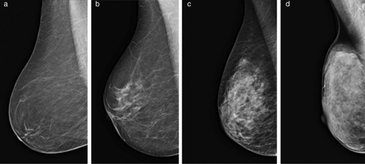
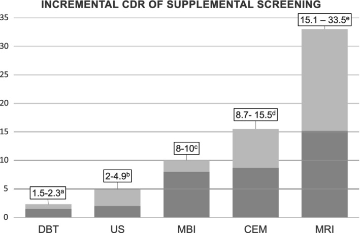
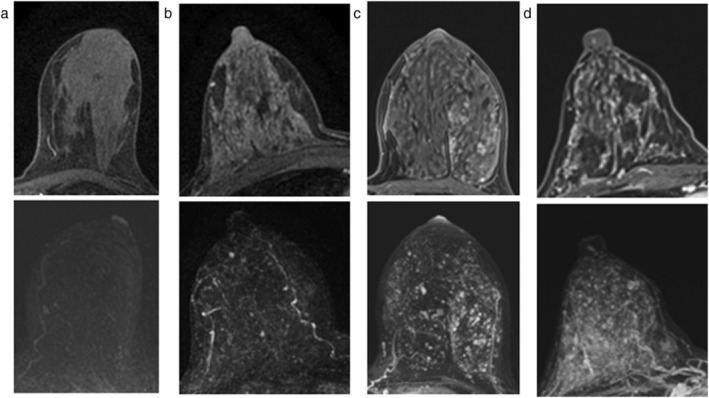
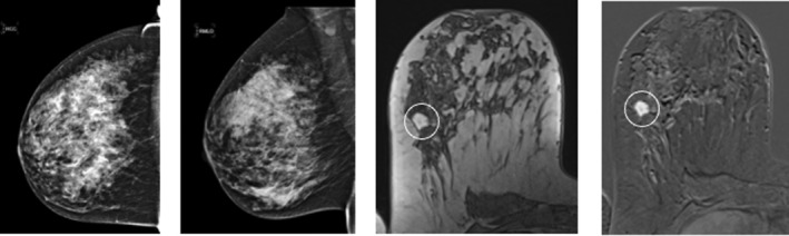
Women with extremely dense breasts are at a higher risk of breast cancer, and the sensitivity of mammography in this group is reduced due to the masking effect of overlapping tissue. This review examines supplemental screening methods to improve detection in this population, with a focus on MRI. Morphologic techniques offer limited benefits, digital breast tomosynthesis (DBT) shows inconsistent results, and ultrasound (US), while improving cancer detection rates (CDR), results in a higher rate of false positives. Functional imaging techniques show better performance, molecular breast imaging increases CDR but is limited in availability, and contrast-enhanced mammography is promising, with good results and as an accessible technique, but requires further validation. MRI, with sensitivity ranging from 81% to 100%, is the most supported modality. Despite strong evidence for MRI in this population, high costs, use of contrast, and longer scan times hinder widespread use. Abbreviated MRI protocols aim to overcome these barriers by reducing costs and scan duration. As personalized screening becomes a future focus, MRI remains the most effective option for women with extremely dense breasts. LEVEL OF EVIDENCE: 1 TECHNICAL EFFICACY: Stage 5.
Keywords: breast density; breast neoplasms; early detection cancer; magnetic resonance imaging.
© 2025 The Author(s). Journal of Magnetic Resonance Imaging published by Wiley Periodicals LLC on behalf of International Society for Magnetic Resonance in Medicine.
Figures

References
-
- McCormack VA, Dos Santos SI. Breast density and parenchymal patterns as markers of breast cancer risk: A meta‐analysis. Cancer Epidemiol Biomarkers Prev 2006;15(6):1159‐1169. - PubMed
-
- D'Orsi CJS, Sickles EA, Mendelson EB, et al. ACR BI‐RADS® Atlas, Breast Imaging Reporting and Data System. 5th ed. Reston, VA: American College of Radiology; 2013.
Publication types
MeSH terms
Grants and funding
LinkOut - more resources
Full Text Sources
Medical
Research Materials
