. 2025 Sep;308(9):2371-2391.
doi: 10.1002/ar.25630. Epub 2025 Jan 24.

Is cranial anatomy indicative of fossoriality? A case study of the mammaliaform Hadrocodium wui

Affiliations

Is cranial anatomy indicative of fossoriality? A case study of the mammaliaform Hadrocodium wui

Molly Tumelty et al. Anat Rec (Hoboken). 2025 Sep.

Abstract

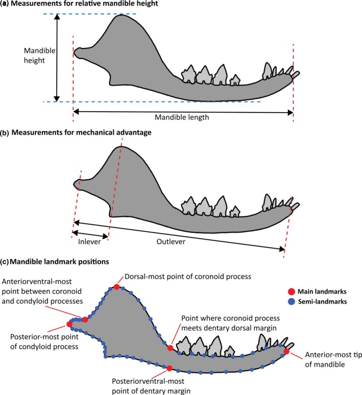
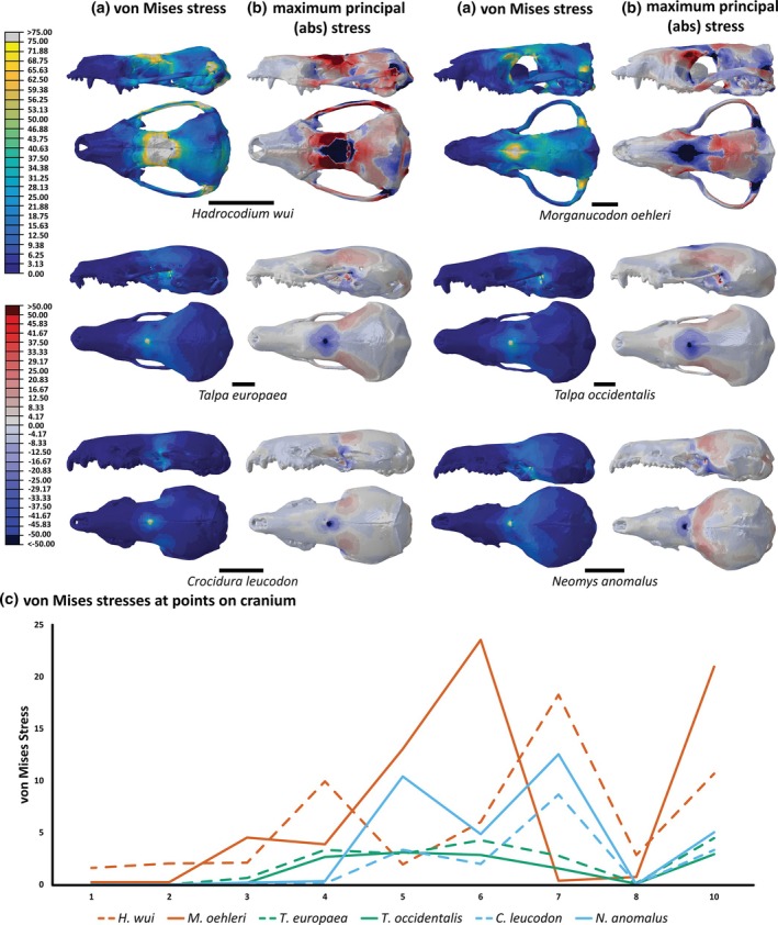
Determining the ecology of fossil species presents considerable challenges due to the often fragmentary preservation of specimens. The mammaliaform Hadrocodium wui from the Jurassic of China is known only from the cranium and mandible but may have had a fossorial lifestyle. It shares morphological similarities with talpid moles and soricid shrews and is closely related to other fossorial mammaliaforms. However, the lack of postcranial elements has so far precluded a definitive assessment regarding its fossorial behavior. Using a combination of geometric morphometric analysis of the lower mandible and finite element analyses of the cranium, comparisons between H. wui and extant groups are made. H. wui resembles talpid moles more closely than shrews in mandible shape. However, there are more similarities between H. wui and semi-fossorial/semi-aquatic moles than fully fossorial moles. The skull of H. wui is particularly weak in every tested biomechanical scenario when compared to the related mammaliaform Morganucodon oehleri and talpid moles. This weakness is potentially a result of the reduction in thickness of the zygomatic arch. In contrast, the shrew crania show similar stress magnitudes and distribution. These results imply that H. wui was not fully fossorial and fed on a diet of softer invertebrates. Skull morphology may therefore not be sufficient to infer fossoriality in forelimb-digging species.

Keywords: finite element analysis; geometric morphometrics; mammaliaformes; mammalian evolution.


Figures

FIGURE 1
FIGURE 1
Phylogenetic context of H. wui and other species discussed in the text. Diet and fossorial adaptations for the fossil species indicated in the phylogeny. Simplified after Lautenschlager et al. (2023).
FIGURE 2
FIGURE 2
Measurements taken on the lower mandible shown on H. wui. (a) Relative mandible height measurements: Mandible length = anterior‐most point of mandible to posterior‐most point of condyloid process; mandible height = dorsal‐most point of coronoid process to ventral‐most point of dentary margin. (b) Mechanical advantage measurements: Outlever = distance between jaw joint and bite point; inlever = distance between jaw joint and muscle attachment point. (c) Landmark placements (red = landmark; blue = semi‐landmark) (Adapted from Morales‐García et al., 2021).
FIGURE 3
FIGURE 3
Models used for each species in FEA in left lateral and dorsal views. Scale bars = 5 mm.
FIGURE 4
FIGURE 4
(a) Positions at which point loads were applied during FEA shown on left lateral view of H. wui. (b) Landmarks on the cranium where stress was measured shown on dorsal view of H. wui: (1) anteriormost point of premaxilla; (2) right lateralmost point of premaxilla; (3) right lateralmost point where premaxilla meets maxilla; (4) right lateralmost point where maxilla meets the jugal; (5) right lateralmost point where zygoma meets squamosal; (6) right lateralmost point of parietal; (7) posteriormost point of parietal; (8) anteriormost point of nasal; (9) central point of frontal; (10) central point of parietal.
FIGURE 5
FIGURE 5
Results for (a) mechanical advantage and (b) relative mandible height for each species. Red = extinct species; gold = golden mole; green = Talpidae; blue = Soricidae; gray = Uropsilinae. See Supplementary Information for further detail.
FIGURE 6
FIGURE 6
PCA plot for mandible shape across all species with convex hulls representing different groups: Green = Talpidae; blue = Soricidae; red = extinct species. Example mandibles shown in respective colors. See Supplementary Information for further detail.
FIGURE 7
FIGURE 7
PCA plot for stress values at each landmark on the cranium (Figure 4b) split into: (a) all loads; (b) parietal load; (c) frontal load; (d) nasal load; (e) teeth load.
FIGURE 8
FIGURE 8
Finite element analysis results for the parietal loads of each species with contour plots of the left lateral and dorsal views of (a) von Mises stress (yellow = higher stress) and (b) absolute maximum principal stress (red = tensile stress; blue = compressive stress. Scale bars = 5 mm. (c) Plot of von Mises stress values at landmarks across cranium.
FIGURE 9
FIGURE 9
Finite element analysis results for the frontal loads of each species with contour plots of the left lateral and dorsal views of (a) von Mises stress (yellow = higher stress) and (b) absolute maximum principal stress (red = tensile stress; blue = compressive stress. Scale bars = 5 mm. (c) Plot of von Mises stress values at landmarks across cranium.
FIGURE 10
FIGURE 10
Finite element analysis results for the nasal loads of each species with contour plots of the left lateral and dorsal views of (a) von Mises stress (yellow = higher stress) and (b) absolute maximum principal stress (red = tensile stress; blue = compressive stress. Scale bars = 5 mm. (c) Plot of von Mises stress values at landmarks across cranium.
FIGURE 11
FIGURE 11
Finite element analysis results for the teeth loads of each species with contour plots of the left lateral and dorsal views of (a) von Mises stress (yellow = higher stress) and (b) absolute maximum principal stress (red = tensile stress; blue = compressive stress. Scale bars = 5 mm. (c) Plot of von Mises stress values at landmarks across cranium.

Similar articles

References

    1. Agrawal, V. C. (1967). Skull adaptations in fossorial rodents. Mammalia, 31(2), 300– 312.
    1. Angielczyk, K. D. , & Schmitz, L. (2014). Nocturnality in synapsids predates the origin of mammals by over 100 million years. Proceedings of the Royal Society B: Biological Sciences, 281(1793), 20141642. - PMC - PubMed
    1. Anthwal, N. , & Tucker, A. S. (2022). Evolution and development of the mammalian jaw joint: Making a novel structure. Evolution & Development, 25(1), 3–14. - PMC - PubMed
    1. Arlton, A. V. (1936). An ecological study of the mole. Journal of Mammalogy, 17(4), 349.
    1. Becerra, F. , Casinos, A. , & Vassallo, A. I. (2012). Biting performance and skull biomechanics of a chisel tooth digging rodent (Ctenomys tuconax; Caviomorpha; Octodontoidea). Journal of Experimental Zoology Part A: Ecological Genetics and Physiology, 319(2), 74–85. - PubMed

LinkOut - more resources