. 2025 May;12(19):e2411540.
doi: 10.1002/advs.202411540. Epub 2025 Jan 24.

Artificial Cornea Substitute Based on Hydrogel Skeletons with Natural Stromal Hierarchical Structure and Extracellular Matrix for Sutureless Transplantation

Affiliations

Artificial Cornea Substitute Based on Hydrogel Skeletons with Natural Stromal Hierarchical Structure and Extracellular Matrix for Sutureless Transplantation

Long Zhao et al. Adv Sci (Weinh). 2025 May.

Abstract

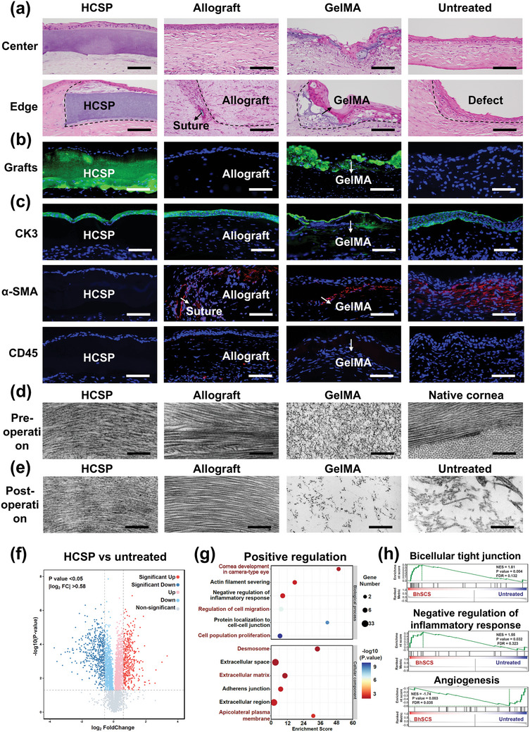
Corneal substitutes with structural and compositional characteristics resembling those of natural corneas have attracted considerable attention. However, biomimicking the complex hierarchical organization of corneal stroma is challenging. In this study, humanized corneal stroma-like adhesive patches (HCSPs) are prepared through a multi-step process. First, polyethylene glycol diacrylate is cast and cured within decellularized porcine cornea (DPC) templates. The DPCs are then enzymatically digested to obtain hydrogel skeletons, which are finally integrated with human corneal extracellular matrix and methacrylate gelatin. HCSPs replicate the ultrastructure, protein components, and optical properties of human corneas and exhibit improved anti-swelling and anti-degradation capabilities compared with conventional DPCs and recombinant human collagen patches. HCSPs can deliver methacrylate gelatin at the ocular surface temperature (37 °C) and achieve stable adhesion to the corneal stroma upon 405 nm light irradiation. Furthermore, HCSPs promote the survival and migration of corneal epithelial and stromal cells while preserving their phenotypes. In rabbit models of lamellar keratoplasty and microperforation repair, HCSPs accelerate epithelial healing, minimize suture-associated complications, and maintain structural stability. These findings suggest that HCSPs are promising donor corneal substitutes for clinical applications.

Keywords: artificial cornea substitutes; biostructure replication; corneal extracellular matrix; hydrogel skeletons; sutureless transplantations.


Conflict of interest statement

The authors declare no conflict of interest.

Figures

Figure 1
Figure 1
Preparation, adhesion mechanism, and biological characteristics of hCECM‐integrated corneal stroma‐like adhesive patch (HCSP). a) Illustration of the anatomical structure of corneal stroma. b) Schematic of human corneal extracellular matrix (hCECM) solution preparation. c) Schematic of inverted matrix fiber nanotubular hydrogel skeleton (IMNS) fabrication. d) Schematic of HCSP construction. e) Adhesion mechanism and applications of HCSP.
Figure 2
Figure 2
Morphology, architecture, and composition of HCSPs. a) Microscopic images showing morphology and macroscopic transparency of HCSPs and human cornea. b) Microscopic images of HCSPs and human corneal stroma obtained after H&E staining; scale bars = 50 µm. c) SEM images of IMNS and free polymerized PEGDA; scale bars = 5 µm. d) SEM images of HCSPs and human corneal stroma; scale bars = 5 µm. e) TEM images of IMNS and free‐polymerized PEGDA. f) TEM images of HCSPs and human corneal stroma. Scale bars of TEM images = 1 µm for low magnification and 200 nm for high magnification. g) Subtypes of matrisome proteins in HCSPs and human corneal stroma. h) Top 20 matrisome proteins in HCSPs. i) Number of core matrisome proteins detected in HCSPs and human corneal stroma.
Figure 3
Figure 3
Physical properties of HCSP. a) Morphology and ultrastructure of HCSPs, DPCs, and NPCs after 0 and 48 h of immersion in artificial tears. (DPC: decellularized porcine cornea; NPC: natural porcine cornea); scale bars = 200 nm for TEM images. Light transmittance of HCSPs, DPCs, and NPCs at b) 0 d and c) 7 d of immersion in artificial tears. d) Swelling ratio. e) Physical states of HCSPs, DPCs, and RHCPs after incubation in collagenase solution for 3 d. For visualization, materials were stained with 0.4% trypan blue. (RHCP: recombinant human collagen patch) f) Percentage of residual mass. g) Results of mechanical strength test. Data represented as mean ± SD. ****, p < 0.0001. ns, not significant.
Figure 4
Figure 4
Tissue adhesion properties of HCSPs. a) Demonstration of temperature and light responsiveness of HCSPs. (i) At 20 °C, red dye‐labeled GelMA within HCSPs did not leach out to stain the model. (ii) At 37 °C, the model surface was stained by exuding GelMA. (iii) After 405 nm light irradiation, HCSPs firmly adhered to the model surface. Rheological properties of HCSPs, 20% GelMA, and IMNS b) at different test temperatures and c) before and after 405 nm light exposure. d) Images showing the adhesiveness of HCSPs to porcine eyeball. e) Adhesion and f) shear strength of HCSPs and commercial adhesives to corneal stroma. (FG: fibrin glue; PEG‐BA: PEG‐based adhesive; CA: cyanoacrylate adhesive). g) Illustration of the burst pressure measurements. Maximum IOP sustained by HCSPs and commercial adhesives at h) 0 h and i) 48 h. Data represented as mean ± SD. *, p < 0.05; **, p < 0.01; ***, p < 0.001; ****, p < 0.0001. ns, not significant.
Figure 5
Figure 5
Cytocompatibility and phenotypic regulation of HCSPs. Representative live/dead fluorescence staining and scratch assay images of a) HCECs and b) HCSCs; scale bars = 100 µm for live/dead staining and 200 µm for scratch assay. c) Survival rate of HCECs and HCSCs. Percentages of repaired areas for d) HCECs and e) HCSCs at 18 and 36 h post scratch. Immunofluorescence images of f) HCECs and g) HCSCs cultured on HCSPs or directly on well plates; scale bars = 100 µm. GO enrichment analysis of upregulated genes in h) HCECs and i) HCSCs (cells cultured on HCSPs versus well plates). Data represented as mean ± SD. ***, < 0.001; ****, p < 0.0001. ns, not significant.
Figure 6
Figure 6
In vivo repair of lamellar stromal defects in rabbit cornea. a) Schematic of procedures for creating lamellar defects and repairing them with HCSPs. b) Slit lamp microscopy and fluorescence staining after stromal excision. c) AS‐OCT images pre‐ and post‐transplantation. Slit‐lamp microscopy and fluorescence staining images of d) HCSP‐, e) allograft‐, and f) 20% GelMA‐repaired groups; and g) untreated controls at 7, 14, and 28 d post‐transplantation. h) AS‐OCT images at 14 and 28 d post‐operation. i) Pachymetry maps obtained pre‐transplantation and at 28 d post‐transplantation.
Figure 7
Figure 7
Histological, ultrastructural, and proteomic evaluations of repaired rabbit corneas. a) H&E staining for HCSP, allograft, and GelMA treated and untreated corneas at 28 d post‐operation; scale bars = 100 µm. b) Fluorescence staining for HCSP‐ and GelMA‐treated corneas at 28 d post‐operation; scale bars = 100 µm. c) Immunofluorescence for CK3, α‐SMA, and CD45 at 28 d post‐operation; scale bars = 100 µm. d) Pre‐implantation TEM images of HCSP, allograft, GelMA, and native cornea; scale bars = 500 nm. e) TEM images of repaired corneal stroma at 28 d post‐operation from various treatment groups; scale bars = 500 nm. f) Volcano plot, g) Significant GO terms for biological processes and cellular components of upregulated proteins, and h) GSEA plots of representative protein sets: bicellular tight junctions, negative regulation of inflammatory response, and angiogenesis (HCSP‐treated vs untreated corneas).
Figure 8
Figure 8
In vivo repairing of rabbit corneal microperforations. a) Schematic showing the creation of corneal perforation and repair using HCSPs. b) Slit‐lamp and fluorescence staining images recorded after corneal perforation. c) AS‐OCT images recorded before and after HCSP transplantation. Slit‐lamp and fluorescence staining images of d) HCSP‐ and e) GelMA‐repaired groups and f) untreated controls recorded at 5, 14, and 28 d post‐transplantation. g) AS‐OCT and h) H&E staining images of native corneas and those in different treatment groups at 28 d post‐operation; scale bars = 100 µm. i) Immunofluorescence staining images of endothelium in native cornea and different treatment groups at 28 d post‐operation; scale bars = 50 µm.

Similar articles

References

    1. Nosrati H., Ashrafi‐Dehkordi K., Alizadeh Z., Sanami S., Banitalebi‐Dehkordi M., Polim. Med. 2020, 50, 57. - PubMed
    1. Whitcher J. P., Srinivasan M., Upadhyay M. P., Bull. World Health Organ. 2001, 79, 214. - PMC - PubMed
    1. Mishra S. K., Joshi A., Ginu P. M., Sati A., Kumar S. V., Med. J. Armed Forces India 2023, 79, 645. - PMC - PubMed
    1. Gain P., Jullienne R., He Z., Aldossary M., Acquart S., Cognasse F., Thuret G., JAMA Ophthalmol. 2016, 134, 167. - PubMed
    1. a) Xu Y., Liu J., Song W., Wang Q., Sun X., Zhao Q., Huang Y., Li H., Peng Y., Yuan J., Ji B., Ren L., Adv. Sci. 2023, 10, 2205878; - PMC - PubMed
    2. b) Kong B., Chen Y., Liu R., Liu X., Liu C., Shao Z., Xiong L., Liu X., Sun W., Mi S., Nat. Commun. 2020, 11, 1435; - PMC - PubMed
    3. c) Majumdar S., Wang X., Sommerfeld S. D., Chae J. J., Athanasopoulou E. N., Shores L. S., Duan X., Amzel L. M., Stellacci F., Schein O., Guo Q., Singh A., Elisseeff J. H., Adv. Funct. Mater. 2018, 28, 1804076; - PMC - PubMed
    4. d) Lei M., Zhang S., Zhou H., Wan H., Lu Y., Lin S., Sun J., Qu X., Liu C., ACS Nano 2022, 16, 10632; - PubMed
    5. e) Kong B., Sun L., Liu R., Chen Y., Shang Y., Tan H., Zhao Y., Sun L., Chem. Eng. J. 2022, 428, 131012.

LinkOut - more resources