. 2025 Jan 28;122(4):e2313978121.
doi: 10.1073/pnas.2313978121. Epub 2025 Jan 24.

Variants in the SOX9 transactivation middle domain induce axial skeleton dysplasia and scoliosis

Affiliations

Variants in the SOX9 transactivation middle domain induce axial skeleton dysplasia and scoliosis

Lianlei Wang et al. Proc Natl Acad Sci U S A. .

Abstract

SOX9 is a crucial transcriptional regulator of cartilage development and homeostasis. Dysregulation of SOX9 is associated with a wide spectrum of skeletal disorders, including campomelic dysplasia, acampomelic campomelic dysplasia, and scoliosis. Yet how SOX9 variants contribute to the spectrum of axial skeletal disorders is not well understood. Here, we report four pathogenic variants of SOX9 identified in a cohort of patients with congenital vertebral malformations. We report a pathogenic missense variant in the transactivation middle (TAM) domain of SOX9 associated with mild skeletal dysplasia and scoliosis. We isolated a Sox9 mutant mouse with an in-frame microdeletion in the TAM domain (Sox9Asp272del), which exhibits skeletal dysplasia including kinked tails, rib cage anomalies, and scoliosis in homozygous mutants. We find that both the human missense and the mouse microdeletion mutations resulted in reduced SOX9 protein stability in cell culture, while Sox9Asp272del mutant mice show decreased SOX9 expression in the growth plate and annulus fibrosus tissues of the spine. This reduction in SOX9 expression was correlated with the reduction of extracellular matrix components, such as tenascin-X and the Adhesion G-protein coupled receptor ADGRG6. In summary, our work identified and modeled a pathologic variant of SOX9 within the TAM domain and demonstrated its importance for SOX9 protein stability. Our work demonstrates that SOX9 stability is important for the regulation of ADGRG6 expression, which is a known regulator of postnatal spine homeostasis, underscoring the essential role of SOX9 dosage in a spectrum of axial skeleton dysplasia in humans.

Keywords: SOX9 (SRY-Box 9); campomelic dysplasia; congenital vertebral malformations; scoliosis; skeletal dysplasia.


Conflict of interest statement

Competing interests statement:The authors declare no competing interest.

Figures

Fig. 1.
Fig. 1.
SOX9 variants identified in four individuals with skeletal dysplasia. (A) A schematic of the SOX9 protein. SOX9 variants identified in this study (green dots), previous studies (orange dots), and the homologs of the mouse variant (the red dot) are labeled. (BE) Sanger sequencing of the four SOX9 variant cases. (F) Clinical presentation of the four patients. Red arrows indicate skeletal malformations. (G) Asp272 and Gly276 are highly conserved residues. DIM: dimerization domain; HMG: high-mobility-group domain; TAM: transactivation middle, PQA: proline, glutamine, and alanine-rich domain; TAC: transactivation C-terminal.
Fig. 2.
Fig. 2.
Effects of the SOX9 variants on DNA-binding and transcriptional activities of SOX9. (A) The EMSA was performed on Cos-7 cells transfected with WT, SOX9 variant mutant proteins, GFP control construct (Control), and 100-fold unlabeled probes (100X) with two sets of DNA probes. The bound dimer and monomer SOX9 proteins are indicated with black and red arrows. (B) The luciferase assay was performed on SOX9 WT and variant proteins using a Col11a2-Luc construct. His165Tyr variant protein was used as a negative control (25) (n = 3 experiments). Bars are plotted with mean +SD. The statistical difference is evaluated by one-way ANOVA followed by Tukey’s multiple comparison test. *P < 0.05, ns: not significant.
Fig. 3.
Fig. 3.
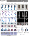
A SOX9 TAM domain mutation in mice leads to axial skeleton defects. (A) Illustration of the in-frame deletion of Asp272del (Sox9del) allele. (BG) Alcian blue and Alizarin red staining of skeletal preparations of WT, Sox9del/+, Sox9del/del, Col2Cre; Sox9flox/+, and Col2Cre; Sox9flox/del mice at P0 (n = 5, 5, 3, 5, and 6, respectively). Magnified views of the forelimb (C), hindlimb (D), thoracic spine I, lumbar spine (L1 and L2) (F), and the palate (G) are provided. Small rib cage, shorter mandible, and campomelia in appendicular bones are indicated with black arrow, red arrow, and red arrowheads in (B). Scapula dysplasia and campomelia in appendicular bones are indicated with orange and red arrowheads in (C and D). Mineralized, unmineralized, and the missing of the T13 floating ribs are indicated with black arrowheads, red arrowheads, and red asterisks in I. Normal or precocious ossification of the vertebrae is indicated with black or red arrowheads in (F). Defects of the palatine and the palatal process of the maxilla are indicated with a red asterisk and red arrowheads in (G). (H) 3D MicroCT renderings of the lumbar spine (L1 and L2) of P0 mice (n = 3). (I) Brightfield images of P5 WT, Sox9del/+, and Sox9del/del mice (n = 12). The kinked tail is pointed by a red arrow. (J and K) X-ray images of WT, Sox9del/+, and Sox9del/del mutant mice at P60 (n = 12 for WT mice; 7 males, 5 females. N = 25 for Sox9del/+ mice; 15 males, 10 females. N = 18 for Sox9del/del mice; 8 males, 10 females). The kinked tail is pointed by a red arrow. The presence or absence of T13 floating ribs is indicated with red arrows or red asterisks. Quantification of the floating rib is shown in (K). (Scale bars, 5 mm in B; 1 mm in CH; and 10 mm in I and J).
Fig. 4.
Fig. 4.
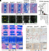
The TAM domain variant results in a region-specific reduction of SOX9 expression in postnatal mouse tails. (A) Alcian Blue Hematoxylin (ABH) staining was performed on the distal regions of the P0 tails of WT, Sox9del/+, Sox9del/del, Col2Cre; Sox9flox/+, and Col2Cre; Sox9flox/del mice (n = 5, 5, 5, 3, and 3, respectively). (B and C) Immunofluorescence (IF) of SOX9 and quantification of SOX9 signal intensity over DAPI were performed on P0 mouse tail sections. (n = 3 for each genotype; 2 vertebrae were analyzed for each mouse.) (D) ABH staining was performed on P5 tail sections of WT, Sox9del/+, and Sox9del/del mice (n = 5). Red and pink arrowheads indicate wedged and unwedged IVDs. The end plate (EP) is indicated with a dotted line and a vertical bar, and the boundary between the inner and outer annulus fibrosus is indicated with a dotted line in the magnified views. (E) IHC analysis of SOX9 on P5 mouse tail sections (n = 5). SOX9 (+) cells are indicated with red arrows. VB: vertebral body; AF: annulus fibrosus; EP: end plate; GP: growth plate; NP: nucleus pulposus; GP-p: proliferative growth plate; GP-h: hypertrophic growth plate; AF-i: inner annulus fibrosus; AF-o: outer annulus fibrosus. (Scale bars, 100 μm.)
Fig. 5.
Fig. 5.
Variants of the TAM domain led to reduced SOX9 protein stability. (A and B) Western blotting and quantification of SOX9 half-life measurements performed on Cos-7 cells transfected with SOX9 WT or variant proteins followed by cycloheximide (CHX) treatment (80 μg/mL) for 0, 1, 2, 4, and 6 h (n = 3 experiments). GAPDH was used as a loading control. Graphs are plotted with mean +SD.
Fig. 6.
Fig. 6.
The TAM domain variant generates global transcriptional changes in chondrocytes and altered protein expression in the spinal tissues. (A) Volcano plot of RNA-sequencing data generated from P5 primary costal chondrocytes of Sox9del/+ and Sox9del/del mice (n = 3 replicas). (B) IF analyses of TNX and SOX9 on P5 tail sections of WT, Sox9del/+, and Sox9del/del mice (n = 5). The nuclei were counterstained with DAPI. (C) IHC analyses of COLI on P5 tail sections of WT, Sox9del/+, and Sox9del/del mice (n = 5). EP: end plate; NP: nucleus pulposus; AF: annulus fibrosus. (Scale bars, 100 μm.)
Fig. 7.
Fig. 7.
Adult Sox9del/del mutant mice display scoliosis and rib cage deformities associated with IVD defects. (A) X-ray images of WT, Sox9del/+, and Sox9del/del mutant mice at 6 mo (n = 8 for WT mice; 4 males, 4 females. n = 15 for Sox9del/+mice; 12 males, 3 females. n = 14 for Sox9del/del mice; 8 males, 6 females.). Magnified sagittal views of the rib cages are provided. Scoliosis and rib cage deformity are indicated with red and yellow arrows. (B), ABH staining was performed on the lumbar spine (L5) at 6 mo (n = 6). Magnified views of the EP and vertebral GP are provided. Disc herniation is indicated with a red arrow. Normal or impaired ossification in the EPs is indicated with black or red asterisks; reduced proteoglycan staining in the GP is indicated with a yellow asterisk. (C and D) IHC analyses of COLX, IHH, and RUNX2 at 6 mo (n = 6). COLX signals in the pericellular matrix and ECM are indicated with red and black arrows. IHH (+) and RUNX2 (+) cells are indicated with red and black arrowheads. (Scale bars, 10 mm in A; 100 μm in BD.)
Fig. 8.
Fig. 8.
A model of the TAM domain variant of SOX9 in perturbing axial skeleton homeostasis. (A) Under physiological conditions, SOX9 maintains normal expression of TNX and COLI in annulus fibrosus and connective tissues and precisely controls the expression of COLX, IHH, and RUNX in EP and GP cells. Mutations in the TAM domain result in reduced SOX9 stability and decreased SOX9 dosage in various spine tissues. (B) The lumbar spine of TAM mutants exhibits IVD degeneration (e.g., disc herniation and increased expression of COLX, IHH, and RUNX2) in the adult stage, associated with late-onset scoliosis. The distal spine (tail), which is more susceptible to SOX9 dosage, displays progressive tail kinking and IVD wedging/degeneration phenotypes in the perinatal and adult stages. Variants in the TAM domain of SOX9 result in reduced expression of some SOX9 targets (e.g., TNX) in tail annulus fibrosus, which may contribute to the postnatal IVD wedging and tail kinking phenotype. This process may be exacerbated with enhanced ossification in the perichondrium and increased cortical bone formation in the tail vertebrae, potentially due to up-regulated IHH expression in the EP and GP. VB: vertebral body; CT: connective tissue; EP: end plate; NP: nucleus pulposus; GP-p: proliferative growth plate; GP-h: hypertrophic growth plate; AF-i: inner annulus fibrosus; AF-o: outer annulus fibrosus; P: perichondrium; CB: cortical bone.

Update of

References

    1. Lefebvre V., Roles and regulation of SOX transcription factors in skeletogenesis. Curr. Top. Dev. Biol. 133, 171–193 (2019). - PMC - PubMed
    1. Sugimoto Y., et al. , Scx+/Sox9+ progenitors contribute to the establishment of the junction between cartilage and tendon/ligament. Development 140, 2280–2288 (2013). - PubMed
    1. Henry S. P., Liang S., Akdemir K. C., de Crombrugghe B., The postnatal role of Sox9 in cartilage. J. Bone Miner. Res. 27, 2511–2525 (2012). - PMC - PubMed
    1. Tsingas M., et al. , Sox9 deletion causes severe intervertebral disc degeneration characterized by apoptosis, matrix remodeling, and compartment-specific transcriptomic changes. Matrix Biol. 94, 110–133 (2020). - PMC - PubMed
    1. Lefebvre V., Dvir-Ginzberg M., SOX9 and the many facets of its regulation in the chondrocyte lineage. Connect Tissue Res. 58, 2–14 (2017). - PMC - PubMed