. 2025 Jan 24;11(4):eadq7706.
doi: 10.1126/sciadv.adq7706. Epub 2025 Jan 24.

ZDHHC2 promoted antimycobacterial responses by selective autophagic degradation of B-RAF and C-RAF in macrophages

Affiliations

ZDHHC2 promoted antimycobacterial responses by selective autophagic degradation of B-RAF and C-RAF in macrophages

Junli Sheng et al. Sci Adv. .

Abstract

S-Palmitoylation is a reversible post-translational modification involving saturated fatty acid palmitate-to-cysteine linkage in the protein, which guides many aspects of macrophage physiology in health and disease. However, the precise role and underlying mechanisms of palmitoylation in Mycobacterium tuberculosis infection of macrophages remain elusive. Here, we found that M. tuberculosis infection induced the expression of zinc-finger DHHC domain-type palmitoyl-transferases (ZDHHCs), particularly ZDHHC2, in mouse macrophages. Furthermore, ZDHHC2 deficiency in mouse macrophages impaired the immunity against M. tuberculosis and reduced the production of various proinflammatory cytokines. Mechanistic studies revealed the involvement of ZDHHC2 in mediating the palmitoylation of B-RAF and C-RAF, affecting their autophagic degradation and stabilizing protein levels. The increased abundance of B-RAF and C-RAF subsequently increases the activity of the extracellular signal-regulated kinase (ERK) signaling pathway, affecting the survival of M. tuberculosis within macrophages. These findings suggest that ZDHHC2 is a potential target for treating tuberculosis.


Figures

Fig. 1.
Fig. 1.. Expression of ZDHHC2 in macrophages induced by M. tuberculosis infection.
(A) Heatmap of ZDHHCs family derived from RNA sequencing (GSE116357) in WT BMDMs that were either not infected (Ctrl) or infected with M. tuberculosis (H37Rv) for 12 hours. (B) Expression map of ZDHHCs family mRNA levels by using qPCR, in WT mouse BMDMs infected with M. tuberculosis for the indicated time. (C) Protein level of ZDHHC2 were detected by Western blot after BMDMs infected with M. tuberculosis for the indicated time (D) Densitometry quantification of (C) by using the ImageJ software. Data are the representative results of three independent experiments and showed as mean ± SD. Nonsignificant (ns) for P > 0.05, *P < 0.05, and **P < 0.01 by using Mann-Whitney U test.
Fig. 2.
Fig. 2.. The deficiency of ZDHHC2 in mice inhibits the immune response against M. tuberculosis.
(A) Model diagram of M. tuberculosis infection in mice. (B and C) shows the log-fold change (Log10) of bacterial burden in the lungs and spleen after M. tuberculosis infected for 7 and 28 days (n = 5 per group). (D and E) IL-1β, IL-6, IFN-β, and TNF-α, expression level in the lungs (D) and serum (E) from the mice described in (A) was measured by ELISA after infection for 7 days. Data are the representative results of three independent experiments and showed as mean ± SD. Nonsignificant (ns) for P > 0.05, *P < 0.05, **P < 0.01, and ***P < 0.001 by using unpaired two-tailed t test.
Fig. 3.
Fig. 3.. ZDHHC2 deficiency in macrophage inhibits anti-TB immune response in vitro.
(A and B) BMDMs were stimulated by M. tuberculosis (MOI = 5) for the indicated time. The mRNA and protein expression of ZDHHC2 were analyzed using qPCR and Western blot. (C) Statistical chart of bacterial load in BMDMs of (A). (D) The expression of mRNA encoding TNF-α, IL-6, IL-1β, and iNOS from the cells described in (A) was assessed using qPCR. (E) The secretion of TNF-α, IL-6, and IL-1β from the cells described in (A) was measured at 0, 12, 24, or 48 hours after infection by ELISA, while the generation of NO was evaluated by Griess assay kits. Data are the representative results of three independent experiments and showed as mean ± SD. Nonsignificant (ns) for P > 0.05, *P < 0.05, **P < 0.01, and ***P < 0.001 by using Mann-Whitney U test (A and D) or unpaired two-tailed t test (C and E).
Fig. 4.
Fig. 4.. ZDHHC2 deficiency in macrophage effects ERK signaling pathway.
(A) Western blot analysis of the phosphorylation status of the indicated proteins in WT and Zdhhc2−/− BMDMs at various time points ranging from 0 to 2 hours following infection with M. tuberculosis (MOI = 5). GAPDH was an internal control. Representative blots are depicted on the left. (B) Densitometry quantification is presented on the right by using the ImageJ software. (C) Western blot analysis of the phosphorylation status of ERK signaling pathway in WT and Zdhhc2−/− BMDMs at various time points ranging from 0 to 2 hours following infection with M. tuberculosis. GAPDH was an internal control. Representative blots are depicted on the left, while part of blots’ densitometry quantification is presented on the right (D). Data are the representative results of three independent experiments and showed as mean ± SD. Nonsignificant (ns) for P > 0.05,*P < 0.05, **P < 0.01, and ***P < 0.001 by using unpaired two-tailed t test.
Fig. 5.
Fig. 5.. ZDHHC2 interacts with B-RAF and C-RAF and promotes their palmitoylation modification.
(A) HEK293T cells were cotransfected with Flag-tagged ZDHHC2, Myc-tagged B-RAF, and Myc-tagged C-RAF plasmids. Cell lysates were coimmunoprecipitated with anti-Flag beads and immunoblotted with anti-Myc antibody. (B) BMDMs were infected by M. tuberculosis for 1 hour and cell lysates were coimmunoprecipitated with either anti–B-RAF antibody or anti–C-RAF antibody and then immunoblotted with anti-ZDHHC2, anti–B-RAF, and anti–C-RAF antibody. (C and E) Streptavidin blot detection of palmitoylated B-RAF or C-RAF in HEK293T cells transfected with indicated plasmids by Click chemistry assays with or without hydroxylamine (NH2OH, 1 M). The S-palmitoylated B-RAF or C-RAF in immunoprecipitated samples was detected using HRP-streptavidin. The three lines of input align with the first three and last three lanes of IP in the upper panel. (D and F) the densitometry quantification of relative palmitoylated B-RAF or C-RAF is presented by using the ImageJ software. (G and H) Click chemistry assays assess the palmitoylation of B-RAF or C-RAF in HEK293T cells transfected with Flag-tagged Zdhhc2 WT or Zdhhc2 C156A plasmids. Data are the representative results of three independent experiments and showed as mean ± SD. *P < 0.05 by unpaired two-tailed t test.
Fig. 6.
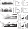
Fig. 6.. The palmitoylation modification of B-RAF and C-RAF can enhance their protein stability.
(A) Immunoblot analysis showing B-RAF/C-RAF degradation in WT BMDM treated with 40 μM CHX for indicated time upon pretreated with dimethyl sulfoxide (DMSO) or 2-BP (100 μM, 12 hours). (B) The densitometry quantification of B-RAF and C-RAF describe in (A) is measured by using the ImageJ software. (C) Immunoblot analysis showing B-RAF/C-RAF degradation in WT or Zdhhc2−/− BMDM after being infected with M. tuberculosis (MOI = 2) and treated with 40 μM CHX for 0, 1, 2, and 4 hours. (D) the densitometry quantification of B-RAF and C-RAF describe in (A) is measured by using the ImageJ software. (E) Western blot showing B-RAF protein expression in HEK293T transfected with Flag-tagged ZDHHC2 or Flag-tagged ZDHHC2C156A as gradient concentrations for 24 hours. (F) the densitometry quantification of B-RAF describe in (E) is measured by using the ImageJ software. (G) Western blot showing C-RAF protein expression in HEK293T transfected with Flag-tagged ZDHHC2 or Flag-tagged ZDHHC2C156A as gradient concentrations for 24 hours. (H) The densitometry quantification of C-RAF in (G) measured by the ImageJ software. Data are the representative results of three independent experiments and showed as mean ± SD. Nonsignificant (ns) for P > 0.05, *P < 0.05, **P < 0.01, and ***P < 0.001 by using unpaired two-tailed t test.
Fig. 7.
Fig. 7.. The palmitoylation modification of B-RAF and C-RAF inhibits their autophagic degradation.
(A) Western blot analysis B-RAF/C-RAF abundance in uninfected and infected M. tuberculosis (MOI = 5) for 1 hours. WT and Zdhhc2−/− BMDMs pretreated with MG132 (20 μM), bafilomycin A1 (BafA1; 0.2 μM), or chloroquine (CQ; 50 μM) for 4 hours. (B) The densitometry quantification of B-RAF and C-RAF describe in (A) is measured by using the ImageJ software. (C) HEK293T cells were cotransfected with B-RAF/C-RAF-Myc and indicated HA-tagged Autophagy-related receptors plasmids. Cell lysates were coimmunoprecipitated with anti-HA beads and immunoblotted with anti-Myc antibody. (D) Western blot showing B-RAF expression in WT and Zdhhc2−/− BMDMs after transfected siOptn siTollip for 24 hours. The densitometry quantification of B-RAF measured by ImageJ software is shown on the right, with values expressed as mean ± SD from three independent experiments. (E) Western blot showing C-RAF expression in WT and Zdhhc2−/− BMDMs after transfected siSqstm1 for 24 hours. The densitometry quantification of C-RAF measured by ImageJ software is shown on the right. (F) Schematic diagram elucidating that ZDHHC2-mediated B-RAF and C-RAF palmitoylation restrain autophagic degradation and activates the ERK signaling pathway to enhance macrophage resistance against M. tuberculosis. Data are the representative results of three independent experiments and showed as mean ± SD. Nonsignificant (ns) for P > 0.05, *P < 0.05, **P < 0.01, and ***P < 0.001 by using unpaired two-tailed t test.

References

    1. Bagcchi S., WHO's Global Tuberculosis Report 2022. Lancet. Microbe 4, E20 (2023). - PubMed
    1. Du X., Sheng J., Chen Y., He S., Yang Y., Huang Y., Fu Y., Lie L., Han Z., Zhu B., Liu H., Wen Q., Zhou X., Zhou C., Hu S., Ma L., The E3 ligase HERC5 promotes antimycobacterial responses in macrophages by ISGylating the phosphatase PTEN. Sci. Signal. 16, eabm1756 (2023). - PubMed
    1. Liu C. H., Liu H., Ge B., Innate immunity in tuberculosis: Host defense vs pathogen evasion. Cell. Mol. Immunol. 14, 963–975 (2017). - PMC - PubMed
    1. Cambier C. J., Takaki K. K., Larson R. P., Hernandez R. E., Tobin D. M., Urdahl K. B., Cosma C. L., Ramakrishnan L., Mycobacteria manipulate macrophage recruitment through coordinated use of membrane lipids. Nature 505, 218–222 (2014). - PMC - PubMed
    1. Jankute M., Cox J. A., Harrison J., Besra G. S., Assembly of the mycobacterial cell wall. Annu. Rev. Microbiol. 69, 405–423 (2015). - PubMed

Substances

LinkOut - more resources