. 2025 Jan 24;11(4):eads2664.
doi: 10.1126/sciadv.ads2664. Epub 2025 Jan 24.

Cryo-EM structure and regulation of human NAD kinase

Affiliations

Cryo-EM structure and regulation of human NAD kinase

Prakash P Praharaj et al. Sci Adv. .

Abstract

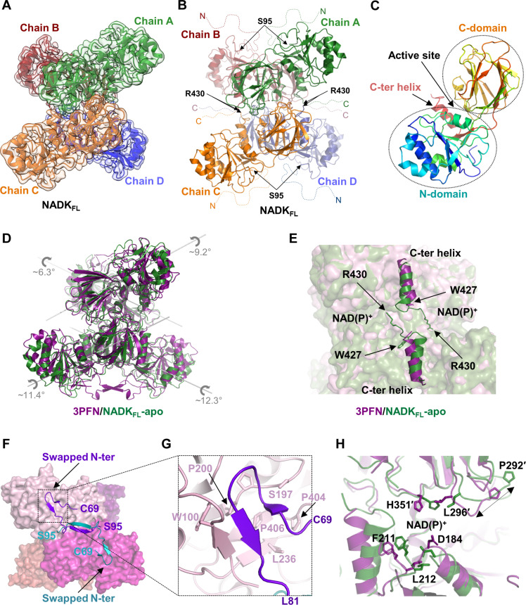
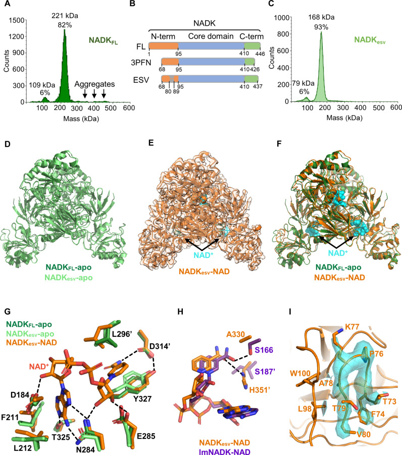
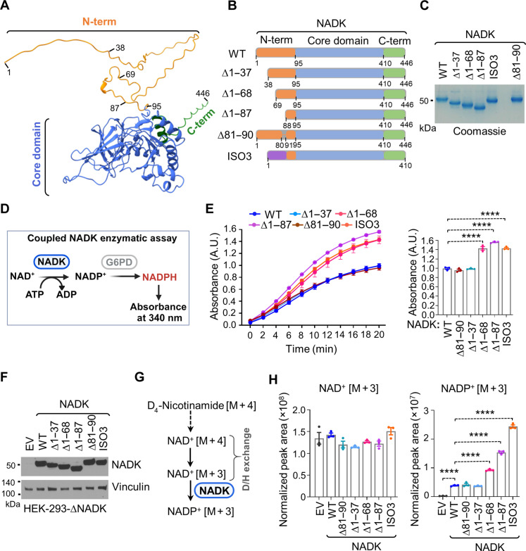
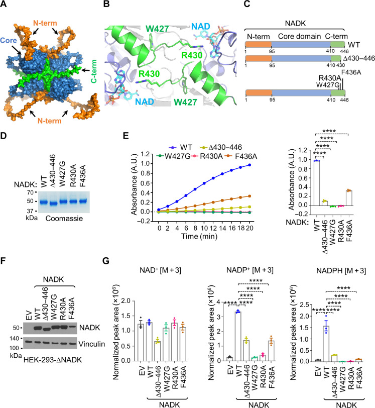
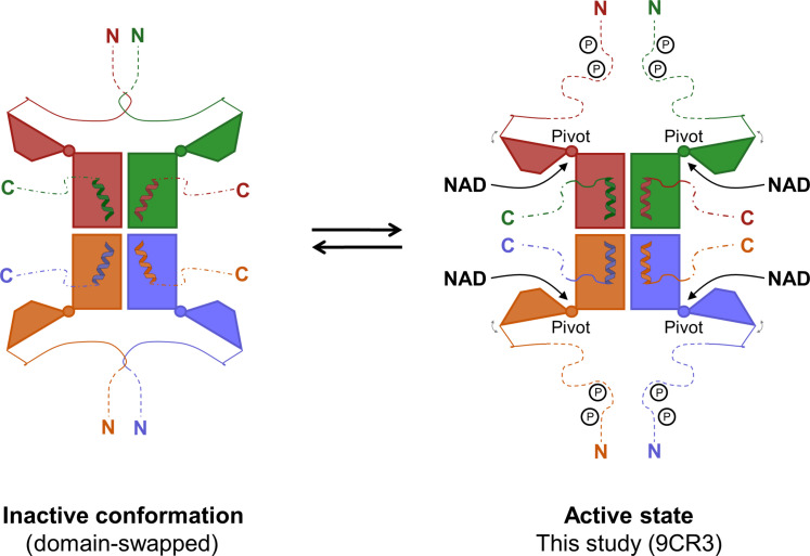
Reduced nicotinamide adenine dinucleotide phosphate (NADPH) is a crucial reducing cofactor for reductive biosynthesis and protection from oxidative stress. To fulfill their heightened anabolic and reductive power demands, cancer cells must boost their NADPH production. Progrowth and mitogenic protein kinases promote the activity of cytosolic NAD kinase (NADK), which produces NADP+, a limiting NADPH precursor. However, the molecular architecture and mechanistic regulation of human NADK remain undescribed. Here, we report the cryo-electron microscopy structure of human NADK, both in its apo-form and in complex with its substrate NAD+ (nicotinamide adenine dinucleotide), revealing a tetrameric organization with distinct structural features. We discover that the amino (N)- and carboxyl (C)-terminal tails of NADK have opposing effects on its enzymatic activity and cellular NADP(H) levels. Specifically, the C-terminal region is critical for NADK activity, whereas the N-terminal region exhibits an inhibitory role. This study highlights molecular insights into the regulation of a vital enzyme governing NADP(H) production.


Figures

Fig. 1.
Fig. 1.. Cryo-EM structure of NADKFL revealing a tetrameric architecture.
(A) Cryo-EM structure of hsNADKFL purified from mammalian cells, revealing a tetrameric organization. Tetrameric NADK is shown embedded within its electron density, with each monomer in a distinct color that are labeled as chains A to D. (B) The organization of the tetrameric hsNADKFL is depicted with each monomer shown in a distinct color as in (A), and their disordered tails are indicated by dashed lines. The boundaries of the catalytic domain visible in the cryo-EM structure are indicated by arrows showing the first (S95) and last (R430) residues, modeled in the density for the N-tail and C-tail, respectively. (C) Domain organization of hsNADKFL, showing the N-terminal domain, the C-terminal domain, the C-terminal helix (residues 413 to 424), and the position of the active site. The monomer is colored in rainbow from its N-terminal (blue) to its C-terminal (red). (D) Comparison of the apo hsNADKFL cryo-EM structure in green and the crystal structure of a truncated construct of hsNADK (PDB: 3PFN) in violet. The rotation axis for each monomer is shown. (E) Comparison of the orientation of the C-terminal helix (residues 413 to 424) in the crystal structure of hsNADK (3PFN) (violet) and the hsNADKFL cryo-EM structure (green). (F) Illustration of the domain swapping for the N-tail (residues 69 to 94) shown in violet and blue in the crystal structure 3PFN. NADK monomers are shown in different shades of pink. (G) Zoom of the contacts between the catalytic domain (pink ribbon) and the swapped N-tail (in violet ribbon). (H) Close-up of the active site of NADK from the 3PFN and cryo-EM structures, showing the indicated structural rearrangements. The black double arrow highlights the translation of P292. (A) to (H) were drawn in PyMOL.
Fig. 2.
Fig. 2.. Cryo-EM structures of the engineered stable variant, hsNADKesv, in apo- and NAD+-bound forms.
(A) Mass photometry of hsNADKFL purified from HEK-293 cells, showing a tetrameric NADK (82%) with some high-molecular-weight aggregates. (B) Schematic of the various constructs of hsNADK (WT; PDB: 3PFN; hsNADKesv). The catalytic domain is shown in blue, the N-tail in orange, and the C-tail in green (including the very C-terminal helix). (C) Mass photometry of hsNADKesv, showing a predominant tetramer (93%). (D) Superimposition of the cryo-EM structures of the apo-forms of hsNADKesv (light green ribbons) and hsNADKFL (dark green ribbon), indicating subtle differences. (E) The cryo-EM structure of the NAD+-bound form of hsNADKesv in orange ribbons and sticks is displayed embedded in its electron density. The NAD+ molecule is shown in cyan. (F) Superimposition of the cryo-EM structures of the apo-form of hsNADKFL (dark green ribbon) and the NAD+-bound form of hsNADKesv (orange ribbons). The NAD+ molecule is shown as cyan spheres. (G) Interactions between hsNADK and the bound NAD+ molecule. Hydrogen bonds are shown as dashed lines. (H) Comparison of the recognition of the amide group of the nicotinamide moiety in hsNADKesv (orange ribbons and sticks) and in the bacterial NADK from Listeria monocytogenes (PDB: 2I29) (violet ribbons and sticks). (I) Electron density of the N-tail: The electron density for the visible part of the N-tail (residues 73 to 80) in the NAD+-bound NADKesv structure is shown as an orange surface and mesh. The backbone and side chains are depicted as sticks, while the rest of the structure is shown as orange ribbons. (D), (F), and (G) were drawn using PyMOL.
Fig. 3.
Fig. 3.. Disordered N-terminal domain inhibits NADK activity and cellular NADP+ biosynthesis.
(A) Ribbon structure of NADK showing the disordered N terminus (1 to 95) in orange, the kinase domain (96 to 429) in blue, and the C-terminal (410 to 446) region in green. PyMOL was used to depict the various regions using the AlphaFold model of human NADK. (B) Schematics of NADK (full length) or various deletion (Δ) variants, including ∆1 to 37, ∆1 to 68, ∆1 to 87, ∆81 to 90, and ISO3. The N-terminal domain is depicted in orange, the kinase domain in blue, and the C terminus in green. (C) Coomassie blue staining of the indicated purified NADK variants from HEK-293 cells. (D) Schematics of the coupled NADK enzymatic assay. The absorbance of NADPH is detected at A340. (E) In vitro enzymatic activity assay for the indicated immunopurified NADK variants (WT, ∆1 to 37, ∆1 to 68, ∆1 to 87, ∆81 to 90, and ISO3). Bar graphs show A340 values after 20 min of the reaction. The absorbance values are indicated as the means ± SEM of biological triplicates. The data are representative of three independent experiments. ****P < 0.001 was calculated with the one-way ANOVA. A.U., arbitrary units. (F) Immunoblotting from NADK-deficient HEK-293 cells expressing either an empty vector (EV) or the indicated NADK variants (WT, ∆1 to 37, ∆1 to 68, ∆1 to 87, ∆81 to 90, and ISO3). (G) Schematics of labeling of NAD+ and NADP+ from 2D4-nicotinamide. (H) Normalized peak areas for newly synthesized NAD+ [M + 3] and NADP+ [M + 3] from NADK-deficient HEK-293 cells expressing either EV or the indicated NADK variants (WT, ∆1 to 37, ∆1 to 68, ∆1 to 87, ∆81 to 90, and ISO3) after 2 hours of labeling with 2,4,5,6-2D-nicotinamide. Data are shown as the means ± SEM of biological triplicates. The data are representative of at least two independent experiments. ****P < 0.001 was calculated with the one-way ANOVA.
Fig. 4.
Fig. 4.. The C terminus is essential for maintaining NADK activity.
(A) AlphaFold model of WT hsNADK as a tetramer. The N-tail, catalytic core, and C-tail of hsNADK are shown in orange, blue, and green, respectively. (B) Zoom in the cryo-EM structure of hsNADKesv. The very C-terminal helix (residues 413 to 424) of the catalytic core and the start of the C-tail (residues 427 to 430) are shown as green ribbons. The NAD+ binding site is highlighted with the NAD+ molecules drawn as sticks. The side chains of W427 and R430 are also shown as sticks. Figure drawn using PyMOL as (A). (C) Schematics of NADK (full length) and different C-terminal variants, including ∆430 to 446, W427G, R430A, and F436A, are color coded as follows: N-terminal domain (orange), the kinase domain (blue), and C terminus (green). (D) Coomassie blue staining of the indicated purified NADK C-terminal variants from HEK-293 cells. (E) In vitro enzymatic activity assay for the indicated immunopurified NADK variants (WT, ∆430 to 446, W427G, R430A, and F436A). Bar graphs show A340 values after 20 min of the reaction. The absorbance values are shown as the means ± SEM of biological triplicates. The data are representative of three independent experiments. (F) Immunoblotting from NADK-deficient HEK-293 cells expressing either EV or the indicated NADK variants (WT, ∆430 to 446, W427G, R430A, and F436A). (G) Normalized peak areas for newly synthesized NAD+ [M + 3], NADP+ [M + 3], and NADPH [M + 3], from NADK-deficient HEK-293 cells expressing either EV or the indicated NADK NADP+ [M + 3], after 2 hours of labeling with 2,4,5,6-2D-nicotinamide. Data are shown as the means ± SEM of biological triplicates. The data are representative of at least two independent experiments. ****P < 0.001 was calculated with the one-way ANOVA.
Fig. 5.
Fig. 5.. Schematic model illustrating the regulation of hsNADK by its terminal regions.
In hsNADK, intertwined pairs of N-terminal tails lock the catalytic domains in an inactive conformation, preventing the proper orientation of the C-terminal helix and the folding of the C-terminal tail over the active site. PTMs, such as phosphorylation, may trigger the separation of the N-tails, enabling the hinge motion of the N-terminal domain to accommodate NAD. Concurrently, the C-terminal helix readjusts to anchor the C-tail, forming a fully functional active site.

References

    1. Pollak N., Dölle C., Ziegler M., The power to reduce: Pyridine nucleotides – Small molecules with a multitude of functions. Biochem. J. 402, 205–218 (2007). - PMC - PubMed
    1. Ju H. Q., Lin J. F., Tian T., Xie D., Xu R. H., NADPH homeostasis in cancer: Functions, mechanisms and therapeutic implications. Signal Transduct. Target. Ther. 5, 231 (2020). - PMC - PubMed
    1. Chandel N. S., NADPH—The forgotten reducing equivalent. Cold Spring Harb. Perspect. Biol. 13, a040550 (2021). - PMC - PubMed
    1. Hoxhaj G., Manning B. D., The PI3K-AKT network at the interface of oncogenic signalling and cancer metabolism. Nat. Rev. Cancer 20, 74–88 (2020). - PMC - PubMed
    1. Pramono A. A., Rather G. M., Herman H., Lestari K., Bertino J. R., NAD- and NADPH-contributing enzymes as therapeutic targets in cancer: An overview. Biomolecules 10, 358 (2020). - PMC - PubMed

MeSH terms

LinkOut - more resources