. 2025 Jan 24;16(1):968.
doi: 10.1038/s41467-024-55690-0.

An essential role for TASL in mouse autoimmune pathogenesis and Toll-like receptor signaling

Affiliations

An essential role for TASL in mouse autoimmune pathogenesis and Toll-like receptor signaling

Laura Lau et al. Nat Commun. .

Abstract

TASL is an immune adaptor that binds to the solute carrier SLC15A4 and facilitates activation of the transcription factor IRF5 during Toll-like receptor (TLR) signaling. Similar to IRF5 and SLC15A4, single nucleotide polymorphisms (SNPs) within TASL have been implicated in increased susceptibility to systemic lupus erythematosus (SLE) in patients. However, the biological function of TASL in vivo and how SLE-associated SNPs increase disease risk is unknown. Here we report that mice deficient in Tasl lack responses to TLR7/9 stimulation and are protected from autoimmune symptoms induced by Aldara or pristane. Loss of Tasl reduces IRF5 phosphorylation and cytokine production in multiple immune cell types but has no effect on other aspects of TLR signaling. Conversely, an SLE-associated TASL risk variant increases TASL protein expression via codon usage, resulting in augmented cytokine production in human cells. Altogether, our study validates the essential function of TASL in TLR signaling and autoimmune pathogenesis.


Conflict of interest statement

Competing interests: All authors were employees of Amgen Inc. at time of studies. L.L, T.A.C, W.O, and P.S.M are current employees of Gilead Inc. R.H. is a current employee of Genentech Inc. A.C. is a current employee of Exelixis. D.N.B., M.J.R., J.H.L., and J.D. are current employees of Amgen Inc.

Figures
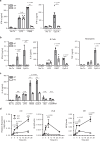
Fig. 1
Fig. 1. Tasl–/– mice show defective response to toll-like receptor 9 and 7 agonists.
a Serum IFN-α levels from Tasl–/– (KO) and wild type (WT) littermate mice injected with either LPS (10 mg/kg), R848 (10 μg), or CpG A (100 μg). n = 5 mice for all treatment groups except WT mice treated with LPS (n = 4). b Splenic plasmacytoid dendritic cells (pDCs), B cells, and neutrophils were isolated from Tasl–/– (KO) and wild type (WT) littermate mice and stimulated with either LPS, R848, or CpG A. Cytokines were measured from supernatants either 24 h post stimulation (pDCs, neutrophils) or 48 h post stimulation (B cells). c Bone marrow derived Flt3L pDCs were stimulated with either poly I:C (50μg/ml), LPS (5 μg/ml), R848 (10 μg/ml), CpG A (10μg/ml), interferon stimulatory DNA ISD (4 μg/ml), or 5’pdsRNA(2 μg/ml) and IL-6 levels was measured from supernatants 24 h post stimulation. d RT-qPCR analysis of Type I IFN genes of WT and KO Flt3L pDCs stimulated with CpG A (1 μg/ml) for the indicated times. All data are means ± SEM and are representative of at least 2 independent experiments. Cells were isolated from 5 mice per genotype that were pooled. In (a), (b), Unpaired t test (2-tailed) *** P < 0.001, ** P < 0.005 In (c) and (d), Two-way ANOVA ***P < 0.0001; ** P < 0.005, *P < 0.05.
Fig. 2
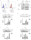
Fig. 2. Tasl–/– mice show defects in IRF5 activation.
a FACS histogram overlays of TLR7 or TLR9 expression from WT and Tasl–/– (KO) mice splenic pDCs. Solid lines indicate staining with primary antibodies, while dotted lines indicate antibody isotype staining controls. b Immunoprecipitation (IP) of TLR7 was performed on Flt3L pDCs derived from WT or Tasl–/– (KO) mice. Western blot analysis was performed for detection of TLR7 processing. HEK293T cells stably expressing mouse TLR7 and TLR4 was used respectively as a positive and negative control. β-actin in whole cell lysate (WCL) was detected as loading control. NS indicates a non-specific band. Data are representative of at least 3 independent experiments c Flt3L pDCs were stimulated with CpG A (3 μM) for the indicated time points. Western blot analysis and quantification was performed for JNK and total JNK and (d) p38 and p-p38. e Western blot analysis and quantification of IRF5 and p-IRF5 in CpG A (3 μM) stimulated Flt3L pDCs at the indicated time points. f Western blot analysis and quantification of p-IRF5 and IRF5 in in CpG A (3μM) stimulated splenic B cells at the indicated time points. Quantification of western blot is represented as the ratio of intensities relative to time zero. n = 5 mice per genotype were used and pooled per experiment.
Fig. 3
Fig. 3. Tasl–/– mice are protected in mouse models of SLE.
Wild type control littermates (WT) and Tasl-/- (KO) mice were treated with either (PBS) or 5% imiquimod (IMQ; Aldara) three times per week and analyzed after 8 weeks. a Representative macroscopic images of spleens. b Mouse spleen weights for treatment groups PBS (n = 5) and IMQ (n = 20). c Serum levels of anti-dsDNA antibodies. PBS (n = 5), IMQ (n = 10). d Representative microscopic images of H&E stained kidney sections. Affected glomeruli are characterized by enlargement and segmental sclerosis with increased hyalinized mesangial matrix (white arrows) and hypertrophy/ hyperplasia of epithelial cells lining Bowman’s capsules (green arrows). e Renal histopathological scores in mice. PBS (n = 5), IMQ (n = 10). f Wild type control littermates (WT) and TASL–/– (KO) mice were injected with either (PBS) (WT n = 5, KO n = 4) or a single dose of TMPD (pristane) (WT n = 15, KO n = 14) and serum anti-nRNP, anti-dsDNA, and anti-Sm autoantibodies were measured 8 months post injection. In (b, c) and (e, f), data are means ± SEM and are representative of 2 independent experiments. Unpaired t test (2 tailed) *** P < 0.0005; ** P < 0.005. Images in (a) and (d) are representative of two independent experiments.
Fig. 4
Fig. 4. SLE associated SNP affects TASL expression.
a DNA and codon triplet sequence of rs887369 alleles for reference (A) and SLE risk variant (C) and codon usage table for Val tRNA frequencies. b HEK293T cells were transfected with either an empty vector (Ctrl) or FLAG tagged TASL expression vectors containing either the rs887369-A SNP or the rs887369-C SNP (risk variant). RT-qPCR analysis of TASL mRNA expression in transfected cells. TASL expression was normalized to neomycin(Neo) resistance cassette co-expressed in the vectors. c Western blot analysis using anti-FLAG (TASL) and β-tubulin (loading control) antibodies. Right panel is quantification of western blot. d Western blot analysis of TASL protein from an in vitro translation assay using FLAG tagged TASL expression vectors containing either the rs887369-A SNP, the rs887369-C SNP (risk variant), or an empty vector (ctrl). N.S. indicates a non-specific protein band. Right panel indicates quantification of TASL bands from two independent experiments normalized to non-specific protein band. e B cells were isolated from PBMCs (n = 3 donors) and were electroporated with Cas9 complexed with either control or gRNAs targeting TASL or MYD88. Cells were stimulated with CpG B and IL-6 was measured 24 h later by CBA. f BLCLs were transfected with Cas9 complexed with either control gRNAs (Parental Ctrl) or gRNAs targeting TASL (KO1, KO2). Cells were stimulated with R848 (10μg/ml) overnight and IL-6 was measured. g human B cells isolated from PBMCs were transfected with mRNA encoding either TASL or CopGFP and stimulated with CpG B. IL-6 was measured 24 h later by CBA. h BLCLs derived from carriers homozygous for either the A or C(risk) rs887369 allele were stimulated with R848 (n = 42). IL-6 was measured from the supernatant 24 h post stimulation. Data are means ± SEM and are representative of at least 2 independent experiments. In (d), (f), (g), and (h) Unpaired t test (2-tailed) **P < 0.01, *P < 0.05. e Two-way ANOVA ***P < 0.001.

References

    1. Weckerle, C. E. & Niewold, T. B. The unexplained female predominance of systemic lupus erythematosus: Clues from genetic and cytokine studies. Clin. Rev. Allergy Immunol.40, 42–49 (2011). - PMC - PubMed
    1. Kirou, K. A. & Gkrouzman, E. Anti-interferon alpha treatment in SLE. Clin. Immunol.148, 303–312 (2013). - PubMed
    1. Isenberg, D. A. B cell targeted therapies in autoimmune diseases. J. Rheumatol. Suppl.77, 24–28 (2006). - PubMed
    1. Baechler, E. C. et al. Interferon-inducible gene expression signature in peripheral blood cells of patients with severe lupus. Proc. Natl. Acad. Sci. USA100, 2610–2615 (2003). - PMC - PubMed
    1. Rowland, S. L. et al. Early, transient depletion of plasmacytoid dendritic cells ameliorates autoimmunity in a lupus model. J. Exp. Med.211, 1977–1991 (2014). - PMC - PubMed

MeSH terms

LinkOut - more resources