. 2025 Jan 24;16(1):978.
doi: 10.1038/s41467-025-56130-3.

SLC25A38 is required for mitochondrial pyridoxal 5'-phosphate (PLP) accumulation

Affiliations

SLC25A38 is required for mitochondrial pyridoxal 5'-phosphate (PLP) accumulation

Izabella A Pena et al. Nat Commun. .

Abstract

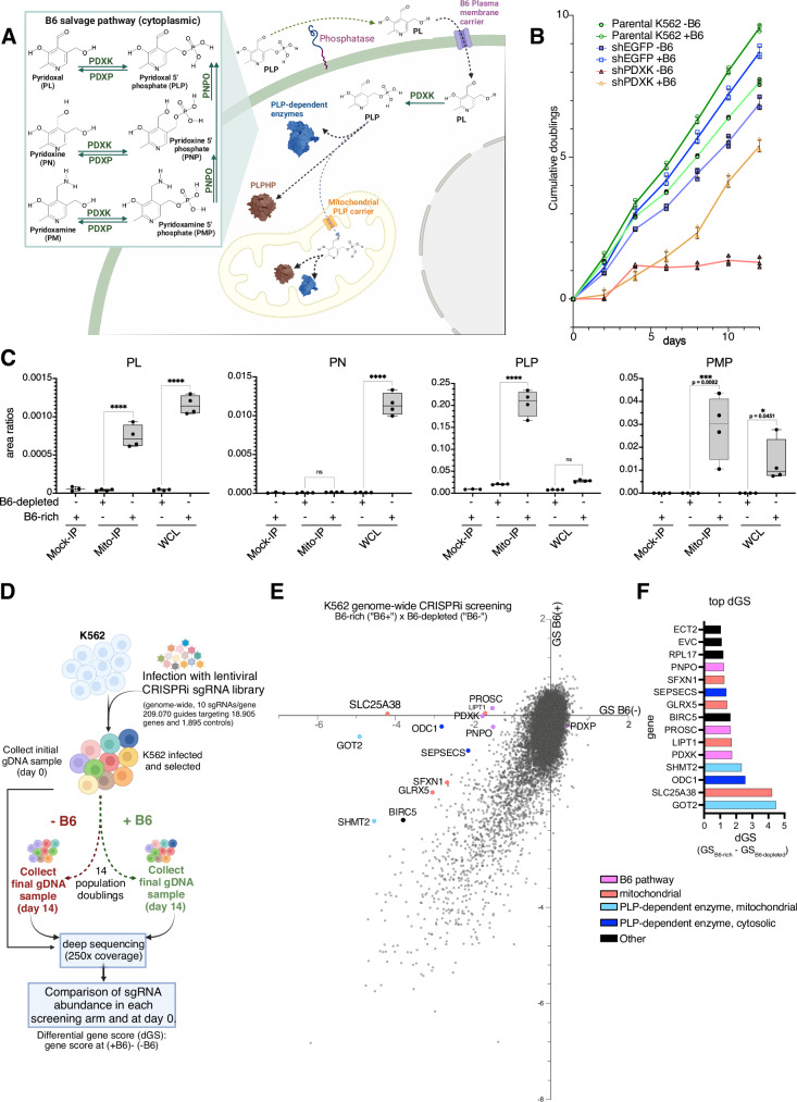
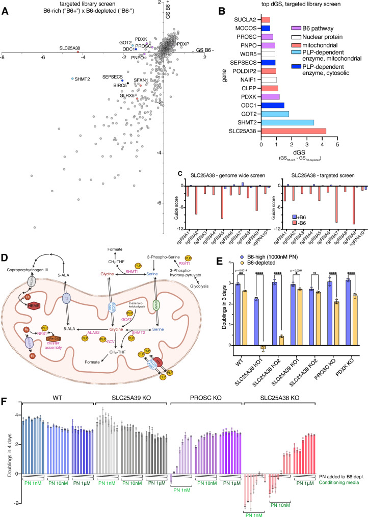
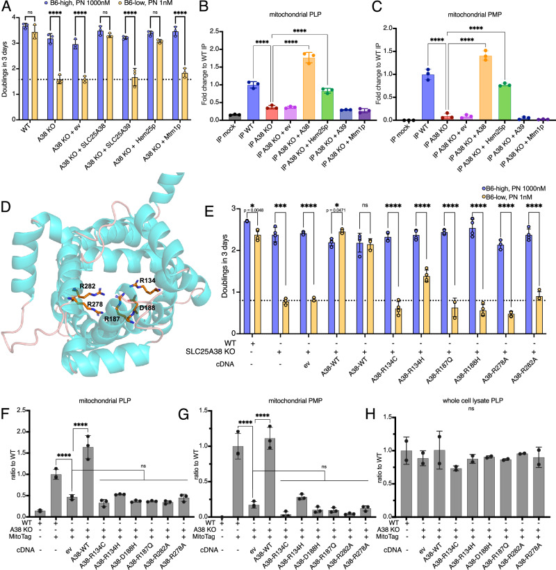
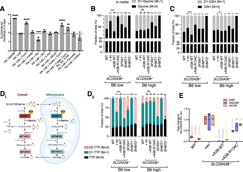
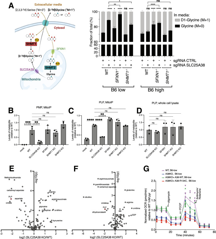
Many essential proteins require pyridoxal 5'-phosphate, the active form of vitamin B6, as a cofactor for their activity. These include enzymes important for amino acid metabolism, one-carbon metabolism, polyamine synthesis, erythropoiesis, and neurotransmitter metabolism. A third of all mammalian pyridoxal 5'-phosphate-dependent enzymes are localized in the mitochondria; however, the molecular machinery involved in the regulation of mitochondrial pyridoxal 5'-phosphate levels in mammals remains unknown. In this study, we used a genome-wide CRISPR interference screen in erythroleukemia cells and organellar metabolomics to identify the mitochondrial inner membrane protein SLC25A38 as a regulator of mitochondrial pyridoxal 5'-phosphate. Loss of SLC25A38 causes depletion of mitochondrial, but not cellular, pyridoxal 5'-phosphate, and impairs cellular proliferation under both physiological and low vitamin B6 conditions. Metabolic changes associated with SLC25A38 loss suggest impaired mitochondrial pyridoxal 5'-phosphate-dependent enzymatic reactions, including serine to glycine conversion catalyzed by serine hydroxymethyltransferase-2 as well as ornithine aminotransferase. The proliferation defect of SLC25A38-null K562 cells in physiological and low vitamin B6 media can be explained by the loss of serine hydroxymethyltransferase-2-dependent production of one-carbon units and downstream de novo nucleotide synthesis. Our work points to a role for SLC25A38 in mitochondrial pyridoxal 5'-phosphate accumulation and provides insights into the pathology of congenital sideroblastic anemia.


Conflict of interest statement

Competing interests: M.G.V.H. discloses that he is an advisor for Agios Pharmaceuticals, iTeos Therapeutics, Sage Therapeutics, Pretzel Therapeutics, Droia Ventures, MPM Capital, and Auron Therapeutics. G.S. is currently an employee and shareholder of AstraZeneca. All other authors declare that they have no competing interests.

Figures

Fig. 1
Fig. 1. A genome-wide CRISPRi screen identifies conditionally essential genes in low vitamin B6 growth conditions.
A Cellular uptake of phosphorylated B6 vitamers involves dephosphorylation by phosphatases like TNSALP. Unphosphorylated forms cross the cell membrane via an unclear mechanism and are phosphorylated by pyridoxal kinase (PDXK). Pyridoxine 5’-phosphate oxidase (PNPO) converts pyridoxine 5’-phosphate (PNP) and pyridoxamine 5’-phosphate (PMP) to pyridoxal 5′-phosphate (PLP) important in the salvage and recycling of intracellular B6. PLPBP (fka PROSC) is key for vitamin B6 homeostasis, possibly a PLP-chaperone. PLP functions as a cofactor for ~60 human enzymes (Supplementary Data 1), forming Schiff bases with lysine residues (catalytic pocket). B Cells grown in media depleted of vitamin B6 display reduced cumulative growth, magnified by PDXK knockdown. C Mitochondria isolated using the “Mito-IP” method and analyzed by LC-MS alongside with the whole cell lysates (“WC”). Mock IP refers to the exact same Mito IP procedure but executed using cells which do not express Mito-tag. Pyridoxal (PL), PLP and PMP were reliably detected in the mitochondrial fraction above the background (Mock IP) but not pyridoxine (PN). PNP and pyridoxamine (PM) signals were below the detection limits of our instrument. Area ratios are shown as peak areas of the target ion divided by peak areas of internal standards (D3-PN and D3-PLP for unphosphorylated and phosphorylated vitamers respectively) normalized by cell counts; Box plots show min. to max. values, median, and SD for n = 4, each an independent culture of cells. Significance level were indicated as *p < 0.05, **p < 0.01, ***p < 0.001, ****p < 0.0001 per two-way ANOVA with Šídák post hoc tests. D Diagram describing the genome-wide CRISPRi screening procedure. E Gene scores (GS) for each gene as identified in the B6 depleted (“B6-“) or B6 rich (“B6+”) arms of the CRISPRi screen are shown, highlighting the highest delta gene-scored (dGS) genes. F Top dGS genes are shown following the described color codes. A created in BioRender. Pena, I. (2025) https://BioRender.com/l99m551. D, created in BioRender. Pena, I. (2025) https://BioRender.com/g50p925. Source data are provided as a Source Data file.
Fig. 2
Fig. 2. Targeted library CRISPRi screen confirms SLC25A38 as a key B6 essential gene in K562 cells and highlights the importance of mitochondrial PLP-dependent metabolism.
A Gene Scores (GS) for the targeted library screen in -B6 or +B6 conditions. B Top differentially essential genes with reduced fitness in B6 stress, with SLC25A38 showing the highest delta Gene Score (dGS: GS in +B6 minus GS in -B6). C Guide scores for SLC25A38 in the genome-wide and targeted CRISPRi screens. D Diagram depicting the PLP-dependent glycine metabolism pathways in mitochondria and cytosol; PLP, pyridoxal 5′-phosphate; CH2-THF, Methylenetetrahydrofolate; 5-ALA, aminolevulinic acid; SHMT, serine hydroxymethyltransferase; GCV, glycine cleavage system (P-protein); ALAS2, Delta-aminolevulinate synthase 2; GCAT, Glycine C-acetyltransferase; PSAT1, phosphoserine aminotransferase 1; SFXN1, sideroflexin 1; ABCB6, ATP-Binding Cassette Sub-Family B Member 6; NFS1, cysteine desulfurase; ISC, iron-sulfur clusters. E Proliferation in B6 rich or B6 poor conditions for clonal knockout cell lines of SLC25A38, SLC25A39, PDXK, and PROSC. Proliferation defects were observed in PDXK, PROSC, and SLC25A38 knockouts in -B6, but not in WT and SLC25A39 knockouts. Cells were plated for proliferation assays after 3 days of conditioning in each respective media. Doublings are shown as mean ± SD; n = 3 independent cell cultures (E), two-way ANOVA followed by Šídák post hoc analysis (E) *p < 0.05, **p < 0.01, ***p < 0.001, ****p < 0.0001. Each n was defined as an independent culture of cells. F K562 cells were conditioned in B6-depleted media supplemented with low B6 (1 nM PN), physiological B6 (10 nM PN), or high B6 (1000 nM PN) for a longer period (6 days, media change every 2 days) and then plated onto 96 well plates at 10.000 cells per well (two replicates) for growth over 4 days in eight media conditions: B6-depleted media without supplementation, +0.5 nM PN, +1 nM PN, +5 nM PN, +10 nM PN, +50 nM PN, +100 nM PN and +1000 nM PN (“PN added to B6-depl.”). Doublings were estimated from absorbance reads using the Presto Blue assay at day 3 (E) or day 4 (F). D Created in BioRender. Pena, I. (2025) https://BioRender.com/m62v944. Source data are provided as a Source Data file.
Fig. 3
Fig. 3. SLC25A38-KO and the yeast mitochondrial glycine transporter Hem25 but not CSA patient variants of SLC25A38 can rescue growth defects in low B6 and mitochondrial PLP levels.
A Growth defect of SLC25A38-KO cells in low B6 is rescued by expression of an add-back construct (FLAG-SLC25A38-WT) and the yeast ortholog, Hem25p (3xMyc tagged). Cells were grown for 3 days in B6-depleted media supplemented with 1 nM or 1000 nM pyridoxine prior to plating 10,000 cells/well in 96 well plates of the respective media with growth accessed using the Presto Blue assay. Low Mitochondrial PLP (B) and PMP (C) levels in SLC25A38-KO cells are also rescued by expression of wild-type SLC25A38 and the yeast ortholog Hem25p but not when SLC25A39 or its yeast orthologue Mtm1p was expressed. D Structural model of the human SLC25A38 highlighting residues in predicted substrate contact sites chosen for mutagenesis studies; asymmetric: R134, R278, and R282; symmetric: R187; amino group binding: R187 and D188. E SLC25A38 point mutant constructs were generated to express mutations affecting putative substrate binding sites: R134C, R143H, R187Q, D188H, R278A, and R282A; expressing these constructs in SLC25A38-KO cells do not fully rescue growth in low B6 but expression of wild-type FLAG-SLC25A38 add-backs (“A38-WT”) does. In line with the lack of growth rescue, SLC25A38-KO expressing the above-mentioned point-mutants fail to rescue mitochondrial PLP (F) and PMP (G) with no significant changes to the whole cell levels of PLP (H). For Mito-IP experiments, cells were grown in media supplemented with 10 nM pyridoxine for 3, days n = 3 ± SD; Doublings are shown for n = 3 ± SD; Two-way ANOVA with Šídák (A, E) and one-way ANOVA (B, C, FH) with Dunnet multiple-comparison tests were used, *p < 0.05, **p < 0.01, ***p < 0.001, ****p < 0.0001. Each n was defined as an independent culture of cells. Source data are provided as a Source Data file.
Fig. 4
Fig. 4. SLC25A38-KO cells fail to accumulate labeled PLP in the mitochondria in physiologically relevant levels of vitamin B6.
A Cells were supplemented with 100 nM of isotopically labeled (2’,2’,2’)-2H3-pyridoxine (D3-PN, “M + 3”) and the deuterated methyl group was traced into mitochondrial D3-PLP by Mito-IP 1 h later. Corrected ion counts are shown for the endogenous unlabeled isotopologue PLP (M0) and labeled, synthesized from the tracer, (2’,2’,2’)-2H3-PLP is shown as D3-PLP (“M + 3”). B Despite detecting a significant signal of M + 3 PLP in the mitochondria of WT cells and SLC25A39-KOs, SLC25A38-KO mitochondria failed to accumulate M + 3 PLP, despite the presence of labeled M + 3 PLP in the whole cell extracts indicating the conversion of M + 3 PN into M + 3 PLP via PDXK and PNPO occurs. Overexpression of WT SLC25A38 (“AB”, add-back) leads to increased import of M + 3 PLP in the mitochondria compared to SLC25A38-KO transduced with an empty vector (“ev) or overexpressing a mutant form of SLC25A38, R134C both after 1 h (C) or 3 h (D) incubation with D3-PN. In both experiments, K562 cells were grown in B6-physiol. medium (10 nM PN) for 2 days prior to the tracing study. Mass isotopomer distributions were corrected for natural abundance using IsoCorrectoR and shown as “corrected ion counts” (BD), n = 3 ± SD; Asterisks indicate significance as in two-way ANOVA Dunnett test for multiple comparison to WT levels (BD), n = 3, *p < 0.05, **p < 0.01, ***p < 0.001, ****p < 0.0001. Each n was defined as an independent culture of cells. Synthetic lethal genetic interactions mined from The Cell Map, for S. cerevisiae hem25Δ (SLC25A38 ortholog (E)), bud17Δ (PDXK ortholog (F)) and tpn1Δ (the yeast pyridoxine transporter in the plasma membrane (G)) and mtm1Δ (SLC25A39 ortholog (H) from genome-wide deletion screens. Genetic interaction scores and statistical confidence measures (p-values) were obtained from The Cell Map, A, created in BioRender. Pena, I. (2025) https://BioRender.com/i81d057. Source data are provided as a Source Data file.
Fig. 5
Fig. 5. SLC25A38-null cells display impairment in de novo glycine synthesis, export, and mitochondrial one-carbon metabolism in low B6.
A Growth defect in B6-low can be rescued by addition of 1 mM formate (Form) but not glycine (Gly), serine (Ser), non-essential amino acids mix (NEAA), folate, 5-aminolevulinic acid (5-Ala), pyruvate or uridine. B Loss of SLC25A38 causes a glycine synthesis defect uniquely in low B6. 2,3,3-2H3-serine was used as a tracer to measure de novo synthesis of 2-2H-glycine and export to the culture media after 12 h using LC-MS. The glycine M + 0 species is the endogenous/unlabeled isotopologue and the glycine M + 1 species (2-2H-glycine) is derived from 2,3,3–2 H3-serine. C Glutathione (GSH) synthesized from glycine via glutathione synthetase is another indirect proxy for glycine levels; GSH M + 1 is derived from the de novo synthesized M + 1 glycine. Di Tracing 2,3,3-2H3-serine into thymidine 5’-triphosphate (TTP) can inform the contribution of cytosolic and mitochondrial one-carbon pathways,,. If 2,3,3-2H3-serine is oxidized by mitochondrial SHMT2 and subsequent enzymes, a singly labeled formate species is formed leading to a one mass unit heavier: M + 1 TTP. In contrast, if 2,3,3-2H3-serine is oxidized by cytosolic SHMT1, a doubly labeled M + 2 TTP is formed. Dii SLC25A38-null cells rely more on cytosolic one-carbon pathways to TTP synthesis uniquely in low B6. SHMT2-null cells can only produce M + 2 TTP and SHMT1-null cells can only produce M + 1 TTP. SFXN1-null cells also display reversal of TTP synthesis towards the cytosolic route, independent of B6 status. E Purine synthesis intermediates GAR (5′-phosphoribosyl-glycinamide), SAICAR (phosphoribosylaminoimidazole-succinocarboxamide) and AICAR (5-aminoimidazole-4-carboxamide ribonucleotide) accumulate in SLC25A38-null, a phenotype rescuable by expression of SLC25A38 WT but not R134C. Box plots show min. to max. values with line at the median. Asterisks indicate significance as in Dunnet post-hoc tests, one-way ANOVA (A, comparing to PN 1 nM, and E, comparing to SLC25A38-KO +ev) and two-way ANOVA (BD, comparisons with WT): *p < 0.05, **p < 0.01, ***p < 0.001, ****p < 0.0001, mean ± SD are shown for n = 3. Each n represents an independent culture of cells. Di, created in BioRender. Pena, I. (2025) https://BioRender.com/y18c741. Source data are provided as a Source Data file.
Fig. 6
Fig. 6. SHMT2 and other mitochondrial PLP-dependent enzymes are affected in SLC25A38-null cells in low B6.
A, left Tracing strategy to investigate if mitochondrially synthesized glycine M + 1 species (2-2H-glycine) can be produced and exported in SLC25A38-null cells in an SHMT2-dependent manner (in SHMT1−/− cells) and whether knocking out SLC25A38 in SFXN1-null cells leads to further impairment of glycine export. A, right Glycine production and export defect is driven by SLC25A38 only in low B6 conditions in WT, SFXN1−/− and SHMT1−/− cells without major effects in supraphysiological B6 (“B6 high”). Genetic block of mitochondrial serine uptake in SFXN1−/− cells or one-carbon metabolism in SHMT1−/− and SHMT2−/− cells does not cause mitochondrial PMP (B) or PLP (C) deficiency nor whole cell PLP changes. D Statistical significance as in Dunnet post-hoc tests comparing to WT levels for two way (A) or one way ANOVA (BD) is shown: *p < 0.05, **p < 0.01, ***p < 0.001, ****p < 0.0001. Mean ± SD is shown for n = 3 in (BD) and n = 2 in (A). HILIC metabolomics reveals a number of amino acid substrates of PLP-dependent enzymes accumulated in the mitochondria of SLC25A38-KO cells compared to WT in K562 (E) and Jurkat (F) (shown here in the log2 of the average fold change; p-value represents two-tailed t-tests, n = 3). PLP was measured using the same mitochondrial IPs by reverse phase chromatography (F5 column) and shown in red, as it cannot be detected using HILIC. G Seahorse analysis using the mitochondrial stress test assay indicates that expression of wild-type FLAG-SLC25A38 (“A38-WT”) but not the R134C point mutant restores oxygen consumption rates (OCR) in B6-low (1 nM PN); n = 6–8, data shown as mean + SEM. Here OCR was normalized by cell count and then fold changes were calculated compared to the WT levels. For all graphs, each n represents an independent culture of cells. A created in BioRender. Pena, I. (2025) https://BioRender.com/a96c224. Source data are provided as a Source Data file.

References

    1. Percudani, R. & Peracchi, A. The B6 database: a tool for the description and classification of vitamin B6-dependent enzymatic activities and of the corresponding protein families. BMC Bioinform.10, 273 (2009). - PMC - PubMed
    1. Wilson, M. P., Plecko, B., Mills, P. B. & Clayton, P. T. Disorders affecting vitamin B6 metabolism. J. Inherit. Metab. Dis.42, 629–646 (2019). - PubMed
    1. van Karnebeek, C., Pena, I. A. & Gospe, S. M. Disorders of pyridoxine metabolism. In (Rosenberg, R. N. & Pascual, J. M. eds.) Rosenberg’s Molecular and Genetic Basis of Neurological and Psychiatric Disease (Sixth Edition) 711–728 (Academic Press, 2020).
    1. Chelban, V. et al. PDXK mutations cause polyneuropathy responsive to pyridoxal 5’-phosphate supplementation. Ann. Neurol.86, 225–240 (2019). - PMC - PubMed
    1. Waymire, K. G. et al. Mice lacking tissue non–specific alkaline phosphatase die from seizures due to defective metabolism of vitamin B–6. Nat. Genet.11, 45–51 (1995). - PubMed

MeSH terms

Substances

Supplementary concepts

LinkOut - more resources