. 2025 Jan 24;15(1):3043.
doi: 10.1038/s41598-025-87844-5.

Genotype-phenotype correlations for 17 Chinese families with inherited retinal dystrophies due to homozygous variants

Affiliations

Genotype-phenotype correlations for 17 Chinese families with inherited retinal dystrophies due to homozygous variants

Xue Fan et al. Sci Rep. .

Abstract

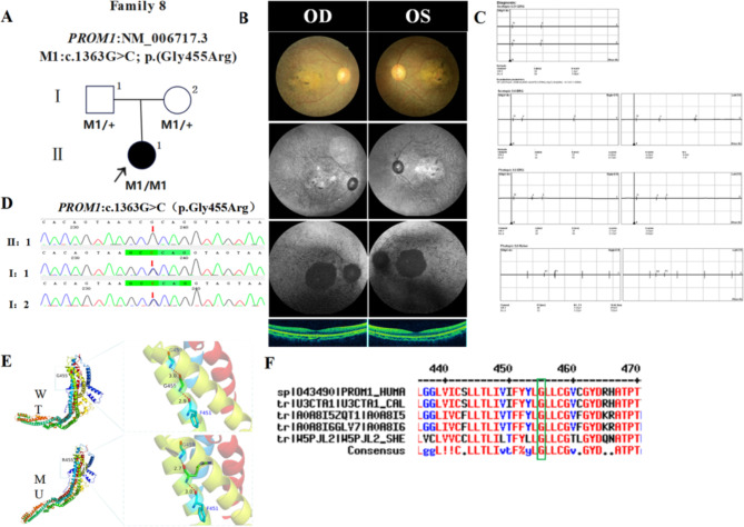
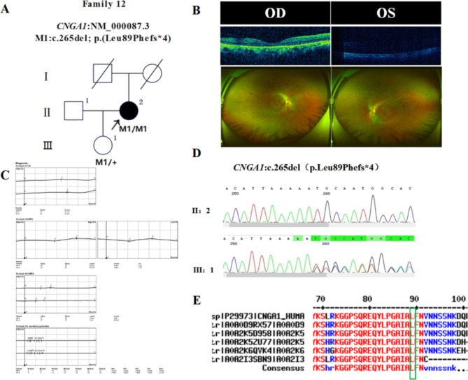
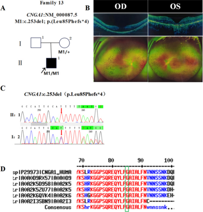
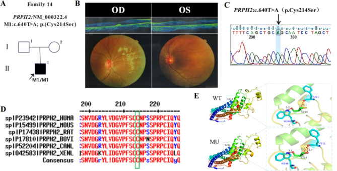
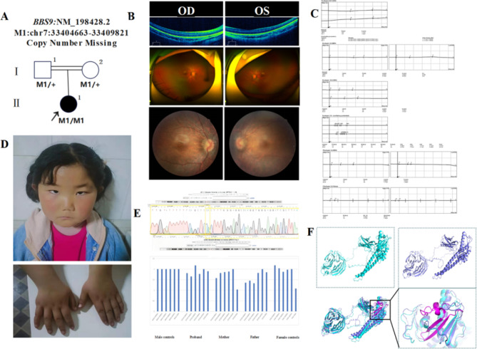
In this study, patients with inherited retinal dystrophies (IRDs) who visited Ningxia Eye Hospital from January 2015 to September 2023 were analyzed. Through Whole Exome Sequencing (WES) and Sanger verification, 17 probands carrying homozygous variants were detected. The association between the genotype and clinical phenotype of patients with homozygous variants was analyzed. Among all the patients, 3 patients (17.6%) had a family history of consanguineous marriage, and the onset age of 5 patients(29.4%) was less than 10 years. According to 12 patients (70.6% ), they had the best corrected visual acuity (monocular) < 0.3. 3 were blind, 9 with moderate to severe visual impairment, and 2 with mild visual impairment. 16 homozygous variants were detected in 9 different genes, of which 7 were novel homozygous variants, including frameshift variants, missense variants, and a copy number variant. These variants are related to clinical phenotypes such as Usher syndrome type II (USH2), Stargardt disease (STGD), retinitis pigmentosa (RP), Leber congenital amaurosis (LCA), and Bardet-Biedl syndrome (BBS) respectively. The results of the study indicate that more than 80% of persons with homozygous variant originated from non-consanguineous families, emphasizing the significance of genetic screening for individuals who lack a family history of consanguineous marriage and no obvious clinical phenotypes, but who may carry genetic pathogenic variants for genetic diseases. Furthermore, by analyzing the genotypes and clinical phenotypes of IRD patients from these 17 Chinese families, we have expanded the spectrum of variants in known pathogenic genes for IRDs and the range of clinical phenotypes associated with variants in these genes. We have identified couples at high risk of having affected offspring and individuals with moderate to severe IRDs, providing a basis for genetic counseling, reproductive decision-making, disease prevention, and management. Our findings highlight the association between homozygous variants and more severe clinical phenotypes within these families, thus laying the groundwork for future genetic screening and intervention strategies.

Keywords: Clinical phenotype; Consanguineous marriage; Genotype; Homozygous variation; Inherited retinal dystrophies.


Conflict of interest statement

Declarations. Competing interests: The authors declare no competing interests. Ethics approval and consent to participate: The Ethics Committee on Human Research at People Hospital in the Ningxia Hui Autonomous Region accepted and examined our work (reference number: 2022-KJCG-006), which adhered to the Declaration of Helsinki. Each participant or their legal guardians provided their written informed permission before to taking part.

Figures

Fig. 1
Fig. 1
variant sequence analysis and clinical examination of the family 4: (A) Pedigree of the family 4: The filled black symbol represents the affected member, and the arrow denotes the proband. (B) Sequence chromatograms of identified variants. (C) The fundus of both eyes: The color of the optic was waxy yellow, the retinal vessels were attenuated, and bone spicule-like pigment deposits were visible on the peripheral retina, OCT showed the disappearance of the light reflection signals in the local ellipsoid zone and interdigitation zone of fovea, along with atrophy of the retinal pigment epithelium. (D) ERG: Significant impairment of cone and rod cell function in both eyes. (E) The homology of amino acid sequences between human USH2A and other species, the amino acid at positions 3393 is highly conserved among species, and the mutated residues 3393 is boxed and indicated.
Fig. 2
Fig. 2
variant sequence analysis and clinical examination of the family 7: (A) Pedigree of the family 7: The filled black symbol represents the affected member, and the arrow denotes the proband. (B) The fundus of both eyes: The color of the optic was pale in both eyes, oval atrophy and yellowish-white patchy exudation were observed in the macular area, showing a bull’s-eye change, OCT indicated a significant thinning of the macular fovea, disappearance of the outer nuclear layer and ellipsoid zone, along with atrophy of the retinal pigment epithelium, fundus angiography reveals ‘worm-eaten-like’ fluorescent spots around the macular region. (C) Sequence chromatograms of identified variants. (D) The homology of amino acid sequences between human PROM1 and other species, the amino acid at positions 182 is highly conserved among species, and the mutated residues 182 is boxed and indicated.
Fig. 3
Fig. 3
variant sequence analysis and clinical examination of the family 8: (A) Pedigree of the family 8: The filled black symbol represents the affected member, and the arrow denotes the proband. (B) The fundus of both eyes: The color of the optic was waxy yellow, the retinal vessels were attenuated, and bone spicule-like pigment deposits were visible on the peripheral retina, OCT showed mild thinning of the macular region. (C) ERG: Significant impairment of cone and rod cell function in both eyes. (D) Sequence chromatograms of identified variants. (E) Proteomic conservation analysis suggested that the p.G455R variant results in the substitution of a nonpolar, uncharged glycine at site 455 with a nonpolar, positively charged arginine, which leads to alterations to the structure and function of the protein after the variant. (F) The homology of amino acid sequences between human PROM1 and other species, the amino acid at positions 455 is highly conserved among species, and the mutated residues 455 is boxed and indicated.
Fig. 4
Fig. 4
variant sequence analysis and clinical examination of the family 12: (A) Pedigree of the family 12:The filled black symbol represents the affected member, and the arrow denotes the proband. (B) The fundus of both eyes: The color of the optic was waxy yellow in both eyes, the retinal vessels were attenuated, and bone spicule-like pigment deposits were visible on the peripheral retina, OCT showed the disappearance of the light reflection signals in the local ellipsoid zone and interdigitation zone at the macular center in the right eye, along with atrophy of the retinal pigment epithelium. (C) ERG: Significant impairment of cone and rod cell function in both eyes. (D) Sequence chromatograms of identified variants. (E) The homology of amino acid sequences between human CNGA1 and other species, the amino acid at positions 89 is highly conserved among species, and the mutated residues 89 is boxed and indicated.
Fig. 5
Fig. 5
variant sequence analysis and clinical examination of the family 13: (A) Pedigree of the family 13:The filled black symbol represents the affected member, and the arrow denotes the proband. (B) The fundus of both eyes: The color of the optic was waxy yellow in both eyes, the retinal vessels were attenuated, and bone spicule-like pigment deposits were visible on the peripheral retina, OCT examination showed the disappearance of the light reflection signals in the local ellipsoid zone and interdigitation zone at the macular center, along with atrophy of the retinal pigment epithelium in the left eye. (C) Sequence chromatograms of identified variants. (D) The homology of amino acid sequences between human CNGA1 and other species, the amino acid at positions 85 is highly conserved among species, and the mutated residues 85 is boxed and indicated.
Fig. 6
Fig. 6
variant sequence analysis and clinical examination of the family 14: (A) Pedigree of the family 14: The filled black symbol represents the affected member, and the arrow denotes the proband. (B) The fundus of both eyes: The color of the optic was pale, OCT showed a mild elevation of the ellipsoid zone and interdigitation zone in the macular region of the the right eye. (C) Sequence chromatograms of identified variants. (D) The homology of amino acid sequences between human PRPH2 and other species. The amino acid at positions 214 is highly conserved among species, and the mutated residues 214 is boxed and indicated. (E) Proteomic conservation analysis suggested that the p.C241S variant results in the substitution of a polar, uncharged cysteine at site 241 with a polar, uncharged serine, which leads to alterations in the structure and function of the mutated protein.
Fig. 7
Fig. 7
variant sequence analysis and clinical examination of the family 17: (A) Pedigree of the family 17: The filled black symbol represents the affected member, double horizontal lines indicate consanguineous marriage, and the arrow denotes the proband. (B) The fundus of both eyes: OCT examination showed no obvious structure of the fovea centralis. (C) ERG: Significant impairment of cone and rod cell function in both eyes. (D) Atient appearance: Epicanthus, wide eye distance, overweight, exotropia, hands have been polydactyly surgery. (E) Breakpoint analysis report and QPCR analysis chart. (F) The overall structural alignment between the mutant and wild-type proteins is quite good, with significant changes observed in the 566–598 structural region of the wild-type and mutant proteins. The mutant is missing two α-helices and two β-sheets, which leads to the shortening of the β-sheets both before and after this region.

Similar articles

References

    1. Scholl, H. P. N. et al. Emerging therapies for inherited retinal degeneration. Sci. Transl Med.8, 368rv6 (2016). - PubMed
    1. Jespersgaard, C. et al. Molecular genetic analysis using targeted NGS analysis of 677 individuals with retinal dystrophy. Sci. Rep.9, 1219 (2019). - PMC - PubMed
    1. Sen, P. et al. Prevalence of retinitis pigmentosa in South Indian population aged above 40 years. Ophthalmic Epidemiol.15, 279–281 (2008). - PubMed
    1. Smith, E. D. et al. Classification of genes: Standardized clinical validity assessment of gene-disease associations aids diagnostic exome analysis and reclassifications. Hum. Mutat.38, 600–608 (2017). - PMC - PubMed
    1. Strande, N. T. et al. Evaluating the clinical validity of gene-disease associations: An evidence-based framework developed by the clinical genome resource. Am. J. Hum. Genet.100, 895–906 (2017). - PMC - PubMed

Publication types

MeSH terms

Supplementary concepts

LinkOut - more resources