. 2025 Jan 24;151(2):45.
doi: 10.1007/s00432-025-06095-z.

Identification of cold tumor induction-related markers in pancreatic cancer and the clinical implication of PCDH7

Affiliations

Identification of cold tumor induction-related markers in pancreatic cancer and the clinical implication of PCDH7

Yuki Mochida et al. J Cancer Res Clin Oncol. .

Abstract

Purpose: Pancreatic ductal adenocarcinoma (PDAC) is considered a "cold" tumor because the tumor immune microenvironment (TIME) exhibits poor intratumoral T-cell infiltration. This study aimed to identify the marker genes associated with induction of cold TIME in PDAC cells.

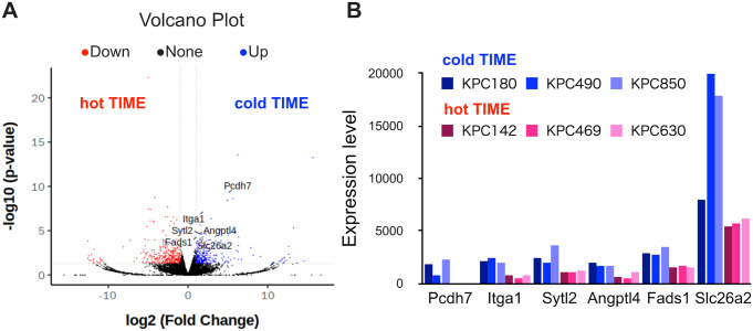
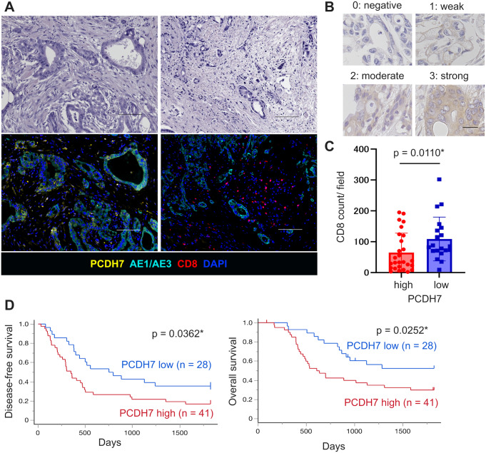
Methods: We orthotopically transplanted 10 primary cultures of PDAC derived from KrasG12D/+; Trp53R172H/+; Pdx-1-Cre (KPC) mice into immunocompetent mice and evaluated TIME by immunohistochemistry (IHC) staining of CD8. We divided primary cultures into two groups: cold TIME group with low CD8+ T-cell infiltration and a hot TIME group with high infiltration. RNA sequencing was performed to identify specific genes in the cold TIME group, and single-cell RNA sequencing (scRNA-seq) data was used for validation. IHC was performed to evaluate expressions in human PDAC samples.

Results: We identified six genes specific in PDAC cells to the cold TIME group by RNA sequencing; these were defined as "cold tumor induction-related genes". Human PDAC scRNA-seq data revealed that cold tumor induction-related genes were significantly and negatively correlated with the number of CD8+ T-cells (p = 0.0341). These genes included protocadherin 7 (PCDH7). High expression of PCDH7 significantly and negatively correlated with the number of CD8+ T-cells in scRNA-seq (p = 0.0474) and IHC (p = 0.0110) data using human PDAC samples. PCDH7 was an independent factor for poor prognosis in PDAC (overall survival: hazard ratio = 2.07, p = 0.0367).

Conclusion: PCDH7 is a prognostic marker associated with CD8+ T-cell infiltration for PDAC patients.

Keywords: CD8-positive T-lymphocytes; PCDH7 protein; Pancreatic cancer; Tumor escape; Tumor microenvironment.


Conflict of interest statement

Declarations. Ethical approval: This study was approved by the Ethics Committee of Kyushu University (approval number: 22002-00). All animal experiments were conducted following the guidelines of the institutional animal committee of Kyushu University (approval number: A22-128 and A23-026). Consent to participate: Written informed consent was waived owing to the retrospective analysis of the study. Competing interests: The authors declare no competing interests.

Figures

Fig. 1
Fig. 1
Identification of KPC mouse–derived primary cultures of PDAC inducing infiltration of CD8+ T-cells in tumors. (A) Representative 3D-culture phase-contrast images of KrasG12D/+; Trp53R172H/+; Pdx-1-Cre (KPC) mouse–derived primary cultures of pancreatic ductal adenocarcinoma (PDAC) with serum or niche factor medium. We classified the morphological features of 3D-cultures based on PDAC differentiation grade. (B) Schematic of the study design. TIME: tumor immune microenvironment; RNA-seq: RNA sequencing. (C) Primary cultures of PDAC cells (2.0 × 105) were transplanted orthotopically, and tumors were resected three weeks later. CD8α immunohistochemistry (IHC) staining images of mouse PDAC tumors (left) and the number of CD8+ T-cells (CD8α-positive cells) per field (right) for each primary culture. Scale bars, 100 μm
Fig. 2
Fig. 2
RNA sequencing identified specific genes inducing cold TIME in KPC mouse–derived primary cultures of PDAC. (A) Volcano plot of cold and hot TIME groups. Red: hot TIME differentially expressed genes (DEGs), blue: cold TIME DEGs (p < 0.05). Cold tumor induction–related genes are labeled. (B) The expression levels of cold tumor induction–related genes in each KPC mouse–derived primary culture of PDAC
Fig. 3
Fig. 3
“Cold tumor induction–related genes” and PCDH7 were associated with cold TIME in human PDAC. (A–C, E and F) Single-cell RNA sequencing (scRNA-seq) analysis of a public database of human PDAC patients. (A) Uniform Manifold Approximation and Projection (UMAP) plot showing the clustering of human PDAC. (B) UMAP plot showing the re-clustering of PTPRC (CD45)-positive immune cells. (C) The correlation between “cold tumor induction–related genes” expression and the number of CD8-positive cells per patient. (D) Kaplan–Meier disease-free survival analysis of cold tumor induction–related genes from The Cancer Genome Atlas (TCGA) pancreatic adenocarcinoma samples (n = 89 in each group). (E and F) The correlation between PCDH7 expression and the percentage of CD8-positive cells (E) and Tregs (F) per patient. (G and H) Using TCGA data of human pancreatic adenocarcinoma patients, the correlation between PCDH7 expression and Treg signature score (G) and immunosuppressive score (H). Spearman’s correlation test or log-rank test was used for analysis. *p < 0.05
Fig. 4
Fig. 4
PCDH7 protein negatively correlated with CD8+ T-cell infiltration in human PDAC patients. (A) Representative hematoxylin and eosin staining images of human PDAC tissues in the upper row and the corresponding immunofluorescence images of PCDH7 (yellow), AE1/AE3 (sky blue), CD8 (red), and DAPI (blue) in the bottom row. Left: high expression of PCDH7 and low number of CD8 + T-cell image. Right: low expression of PCDH7 and high number of CD8 + T-cell image. Scale bars, 100 μm. (B) Representative images of IHC staining for PCDH7. The intensity score: 0, negative; 1, weak; 2, moderate; and 3, strong. Scale bar, 20 μm. (C) Quantification of the CD8α-positive cell number in PCDH7 low and high cancer cells. (D) Disease-free survival and overall survival analysis (performed using Kaplan–Meier analysis) of PCDH7 (high = 41, low = 28) in human PDAC samples. *p < 0.05

References

    1. Ajina R, Weiner LM (2020) T-Cell immunity in pancreatic Cancer. Pancreas 49:1014–1023. 10.1097/MPA.0000000000001621 - PMC - PubMed
    1. Bear AS, Vonderheide RH, O’Hara MH (2020) Challenges and opportunities for Pancreatic Cancer Immunotherapy. Cancer Cell 38:788–802. 10.1016/j.ccell.2020.08.004 - PMC - PubMed
    1. Binnewies M, Roberts EW, Kersten K et al (2018) Understanding the tumor immune microenvironment (TIME) for effective therapy. Nat Med 24:541–550. 10.1038/s41591-018-0014-x - PMC - PubMed
    1. Bolger AM, Lohse M, Usadel B (2014) Trimmomatic: a flexible trimmer for Illumina sequence data. Bioinformatics 30:2114–2120. 10.1093/bioinformatics/btu170 - PMC - PubMed
    1. Chen DS, Mellman I (2013) Oncology meets immunology: the Cancer-Immunity cycle. Immunity 39:1–10. 10.1016/j.immuni.2013.07.012 - PubMed

MeSH terms