Review
. 2025 Jan 24;23(1):45.
doi: 10.1186/s12964-024-02018-6.

Glutamine and cancer: metabolism, immune microenvironment, and therapeutic targets

Affiliations
Review

Glutamine and cancer: metabolism, immune microenvironment, and therapeutic targets

Ding Nan et al. Cell Commun Signal. .

Abstract

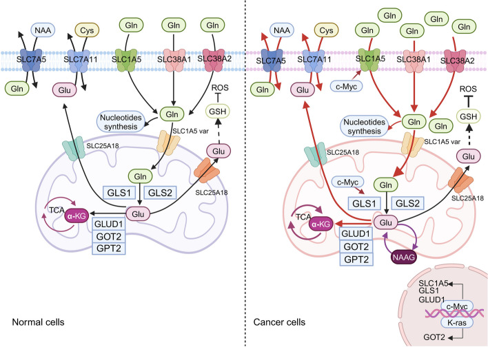
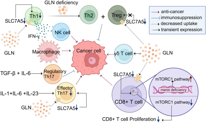
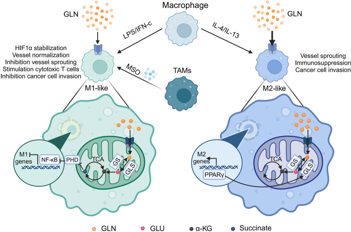
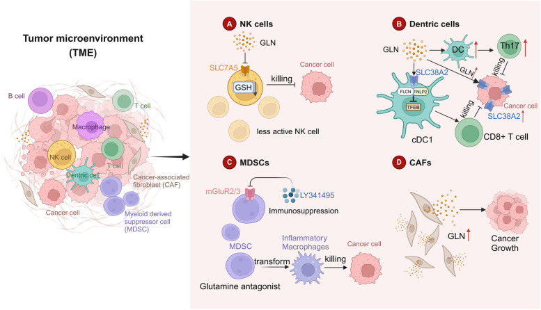
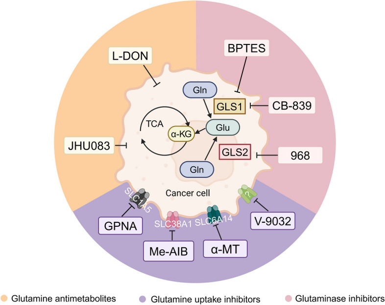
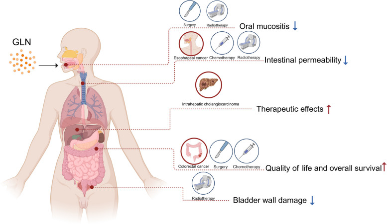
Glutamine is the most abundant amino acid in human serum, and it can provide carbon and nitrogen for biosynthesis, which is crucial for proliferating cells. Moreover, it is widely known that glutamine metabolism is reprogrammed in cancer cells. Many cancer cells undergo metabolic reprogramming targeting glutamine, increasing its uptake to meet their rapid proliferation demands. An increasing amount of study is being done on the particular glutamine metabolic pathways in cancer cells.Further investigation into the function of glutamine in immune cells is warranted given the critical role these cells play in the fight against cancer. Immune cells use glutamine for a variety of biological purposes, including the growth, differentiation, and destruction of cancer cells. With the encouraging results of cancer immunotherapy in recent years, more investigation into the impact of glutamine metabolism on immune cell function in the cancer microenvironment could lead to the discovery of new targets and therapeutic approaches.Oral supplementation with glutamine also enhances the immune capabilities of cancer patients, improves the sensitivity to chemotherapy and radiotherapy, and improves prognosis. The unique metabolism of glutamine in cancer cells, its function in various immune cells, the impact of inhibitors of glutamine metabolism, and the therapeutic use of glutamine supplements are all covered in detail in this article.

Keywords: Cancer; Glutaminase inhibitors; Glutamine antimetabolites; Glutamine metabolism; Immune cells.


Conflict of interest statement

Declarations. Competing interests: The authors declare no competing interests.

Figures

Fig. 1
Fig. 1
Metabolism of glutamine between normal cells and cancer cells. The glutamine transporters (SLC1A5, SLC38A1/SLC38A2, and SLC6A14) expressed on the cell membrane facilitate the transport of glutamine into the cytoplasm. Subsequently, SLC1A5 variants transport glutamine into the mitochondrial matrix, where it is converted to glutamate by GLS. Then, mitochondrial glutamate is converted to α-KG by GLUD1 or various mitochondrial transaminases (including GPT2 and GOT2). Glutamine-derived α-KG provides metabolites for the tricarboxylic acid cycle and can also support OXPHOS or reductive carboxylation pathways. Cancer cells uptake a significant amount of glutamine from plasma through protein transporters. The first and rate-limiting step of glutamine breakdown is catalyzed by GLS in the mitochondria, converting glutamine to glutamate. This enzyme exists in two forms: GLS1 and GLS2, both of which convert it into glutamate. Glutamate is further converted to α-KG by mitochondrial GLUD1 and releases ammonia. α-KG enters the TCA cycle in cancer cells and is converted back to glutamate. In cancer cells, the production of NAAG, derived from glutamine, is significantly increased, and high levels of NAAG are observed in advanced cancer. The NAAG storage cycle provides a mechanism for cancer cells to store this important metabolic product by converting glutamate generated from glutamine into NAAG and then back to glutamate. c-Myc promotes glutamine uptake by targeting the expression of glutamine transporters SLC1A5, SLC38A1, and SLC7A5. Glutamine activates mTORC1 through the SLC7A5/SLC3A2 antiporter system, leading to the efflux of glutamine out of the cell in exchange for leucine influx, a potent Rag-dependent mTORC1 activator. GLS, glutaminase; αKG, α-ketoglutarate; GLUD1, glutamate dehydrogenase 1; GOT, glutamate oxaloacetate transaminase; GPT, glutamate pyruvate transaminase; GSH, glutathione; ROS, reactive oxygen species; OXPHOS, oxidative phosphorylation; GLS1, kidney-type glutaminase; GLS2, liver-type glutaminase; NAAG, N-Acetyl-aspartyl-glutamate
Fig. 2
Fig. 2
Metabolism of glutamine between different kinds of T cells and cancer cells. Th1 combat tumors by secreting IFN-γ to activate macrophages and NK cells. Th2 and Treg promote cancer-induced immune suppression. A mixture of cytokines that bind to glutamine-restricted cells can lead to a shift from Th1 to Th2 expression. Th17 cells can either support or inhibit cancer progression depending on the context. Glutamine metabolism is also a critical regulator of γδ T cell differentiation and IL-17 production. Loss of SLC7A5 results in reduced mTORC1-dependent Th1 and Th17 effector T cells, while Treg numbers are largely unaffected. Proliferation of antigen-stimulated CD8 + T cells already present in the body is completely inhibited. This mechanism is due to the inability of the mTORC1 signaling pathway to activate in the absence of SLC7A5. Furthermore, the absence of the tumor suppressor Menin can enhance mTORC1 signaling and glutamine breakdown. Th1, CD4 + T helper 1 cells; Th2, CD4 + T helper 2 cells; Treg, regulatory T cells; Th17, CD4 + T helper 17; IL-17, interleukin-17
Fig. 3
Fig. 3
The role of glutamine in Macrophages. Macrophages can polarize into two distinct states, M1 and M2. They can be induced by various stimuli, most notably polarizing towards M1 macrophages under LPS/IFN-c stimulation, and towards M2 macrophages under IL-4/IL-13 stimulation. M1 and M2 macrophages have different biological functions. The ratio of succinate to α-ketoglutarate regulates M1 polarization through PHD-dependent proline hydroxylation of IKKβ, which is crucial for NF-κB signaling activation. High levels of succinate in the early stages of M1 polarization favor a strong inflammatory response. Compared to M1 macrophages, M2 macrophages exhibit intact TAC and enhanced OXPHOS. Glutamine itself can influence macrophage phenotype through unknown mechanisms. Another important factor in alternative activation is PPAR-γ signaling. PPAR-γ is not only involved in the conversion of glutamine metabolism but also participates in the upregulation of OXPHOS. TAMs are a special type of macrophage with functional plasticity. MSO regulates TAMs' glutamine metabolism, inducing them towards an M1 phenotype. LPS, lipopolysaccharide; IFN-c, interferon-c; IL-4, interleukin-4; IL-13, interleukin-13; TAC, tricarboxylic acid cycle; OXPHOS, oxidative phosphorylation; TAMs, tumor-associated macrophages; MSO, L-glutamine sulfoximine
Fig. 4
Fig. 4
The role of glutamine in NK cells, dendritic cells, MDSCs and CAFs. A In the TME glutamine is primarily concentrated in cancer cells, leading to a decrease in glutamine levels in NK cells. This reduction in glutamine results in a decrease in GSH levels in NK cells, reducing their cytotoxic function and allowing cancer cells to evade immune attack. B Elevated glutamine levels in cancer patients affect Th17 cell responses by activating the GluR4 receptor on DCs, leading to cancer cell proliferation and tumor growth. Both cDC1s and cancer cells express SLC38A2, with higher expression levels in cancer cells. Mechanistically, in cDC1s, glutamine induces the assembly of the FLCN-FNIP2 complex, inhibiting the activity of the TFEB transcription factor and promoting the antigen-presenting ability of cDC1s. Therefore, glutamine-dependent signaling in cDC1s enhances the generation of cytotoxic effector CD8 + T cells in the TME. C Inhibiting the glutamine metabolism of MDSCs can lead to cell death induced by MDSC activation and their transformation into inflammatory macrophages. mGluR2/3 are expressed on MDSCs, and the mGluR2/3 antagonist LY341495 can weaken the immunosuppressive activity of MDSCs. D CAFs upregulate both glutamine synthesis and secretion, increasing the concentration of glutamine in the TME. TME, tumor microenvironment; NK cells, Natural killer cells; GSH, glutathione; DCs, dendritic cells; cDC1s, type-1 conventional DCs; Th17,CD4 + T helper 17; MDSCs, myeloid-derived suppressor cells;, mGluR2/3, metabotropic glutamate receptors; CAFs, cancer-associated fibroblasts
Fig. 5
Fig. 5
Three main types of drugs that inhibit glutamine metabolism. 1. Glutamine antimetabolites: L-DON can block glutamine-mediated induction of PPAT and PAICS as well as decrease the activity of pyruvate kinase, but it lacks selectivity for glutamine-dependent cells. JHU083 is the prodrug form of L-DON, which can deplete glutamine metabolism in cancer cells, but it can stimulate T-cell proliferation. 2. Glutaminase inhibitors: BPTES is a selective inhibitor of GLS1; CB-839 (telaglenastat) is a selective inhibitor of GLS1 currently undergoing comprehensive clinical trials; 968 is a pan-glutaminase inhibitor, with fourfold selectivity for GLS2 over GLS. 3. Glutamine uptake inhibitors: GPNA can inhibit the growth of colorectal cancer cells by suppressing the overexpressed glutamine transporter SLC1A5 on the cell surface. A GPNA derivative, V-9302, has been developed to effectively inhibit cellular glutamine uptake, but its mechanism of action requires further investigation. MeAIB targeting SLC38A1 and/or SLC38A2, has shown anticancer effects in various cancer cells. α-MT inhibits the growth of SLC6A14-positive breast cancer, PDAC, and CRC cells. L-DON, 6-diazo-5-oxo-L-norleucine; JHU083, ethyl 2-(2-Amino-4-methylpentanamido)DON; BPTES, bis-2-(5-phenylacetamido-1,3,4-thiadiazol-2-yl)ethyl sulfide; GPNA, L-γ-glutamyl-p-nitroanilide; CB-839, telaglenastat; V-9302, 2-amino-4-bis (aryloxy benzyl) aminobutanoic acid; MeAIB, n-methyl-aminoisobutyric acid; α-MT, α-methyltryptophan; PDAC, pancreatic ductal adenocarcinoma; CRC, colorectal cancer
Fig. 6
Fig. 6
The benefits of glutamine supplements in patients with cancer. Supplementation of glutamine has multiple benefits for cancer patients. It can prevent bladder wall damage caused by radiotherapy, possibly through the regulation of extracellular matrix density and collagen expression by glutamine. It can also reduce the incidence of oral mucositis in the subgroup of head and neck cancer patients undergoing radiotherapy. Additionally, it protects lymphocytes in esophageal cancer patients during radiation and chemotherapy and reduces intestinal permeability. It can decrease tumor size and cancer-related cachexia in ICC patients. Furthermore, it improves cellular and humoral immune function in colorectal cancer patients after surgery, while supplementation with L-prolyl-L-glutamine dipeptide can enhance the quality of life and overall survival in colon cancer chemotherapy patients

Similar articles

Cited by

References

    1. Bernfeld E, Foster DA. Glutamine as an Essential Amino Acid for KRas-Driven Cancer Cells. Trends Endocrinol Metab. 2019;30(6):357–68. 10.1016/j.tem.2019.03.003. - PubMed
    1. Yoo HC, Yu YC, Sung Y, Han JM. Glutamine reliance in cell metabolism. Exp Mol Med. 2020;52(9):1496–516. 10.1038/s12276-020-00504-8. - PMC - PubMed
    1. Yang L, Venneti S, Nagrath D. Glutaminolysis: a hallmark of cancer metabolism. Annu Rev Biomed Eng. 2017;19:163–94. 10.1146/annurev-bioeng-071516-044546. - PubMed
    1. “On the Origin of Cancer Cells | Science.” Accessed: Aug. 24, 2023. [Online]. Available: https://www.science.org/doi/10.1126/science.123.3191.309?url_ver=Z39.88-.... - DOI
    1. Ward PS, Thompson CB. Metabolic reprogramming: a cancer hallmark even warburg did not anticipate. Cancer Cell. 2012;21(3):297–308. 10.1016/j.ccr.2012.02.014. - PMC - PubMed

LinkOut - more resources