. 2025 Jan 25;56(1):20.
doi: 10.1186/s13567-025-01449-4.

Binding and cleavage of pro-urokinase by a tegument extract of Fasciola hepatica newly excysted juveniles activate the host fibrinolytic system

Affiliations

Binding and cleavage of pro-urokinase by a tegument extract of Fasciola hepatica newly excysted juveniles activate the host fibrinolytic system

Judit Serrat et al. Vet Res. .

Abstract

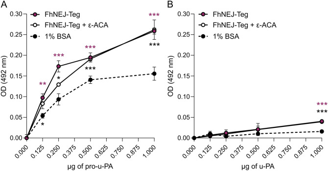
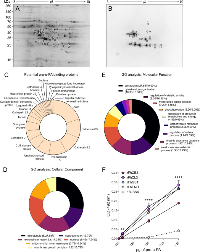
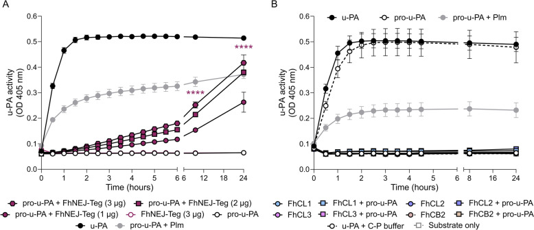
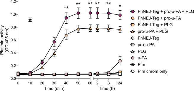
Plasmin, the final product of fibrinolysis, is a broad-spectrum serine protease that degrades extracellular matrix (ECM) components, a function exploited by multiple pathogens for dissemination purposes. The trematode Fasciola hepatica is the leading cause of fasciolosis, a major disease of livestock and an emerging zoonosis in humans. Infection success depends on the ability of F. hepatica newly excysted juveniles (FhNEJ) to penetrate the host intestinal wall, a process that remains incompletely understood. We have previously shown that FhNEJ are capable of binding plasminogen (PLG), the zymogen of plasmin, on their tegument surface, which leads to plasmin generation in the presence of host-derived PLG activators and subsequent degradation of laminin, a major component of the intestinal ECM. Here, we describe the interaction between a tegument extract of FhNEJ and the precursor of the urokinase-type PLG activator (pro-u-PA). We found that F. hepatica cathepsins B3, L3, enolase and glutathione S-transferase mediate this interaction, suggesting a multifactorial or moonlighting role for these proteins. Additionally, our results revealed that the tegument of FhNEJ contains a protease that is capable of cleaving and activating pro-u-PA into its catalytically active form, which positively impacts the capacity of the parasites to generate plasmin from the host PLG. Collectively, our findings indicate that FhNEJ interact with the host fibrinolytic system at multiple levels, reinforcing the potential of targeting this interaction as a strategy to prevent FhNEJ trans-intestinal migration and infection success.

Keywords: Fasciola hepatica; fibrinolytic system; host‒parasite interactions; newly excysted juveniles; pro-urokinase.


Conflict of interest statement

Declarations. Competing interests: The authors declare that they have no competing interests.

Figures

Figure 1
Figure 1
Binding of u-PA and its precursor by FhNEJ-Teg proteins. A, B Binding of FhNEJ-Teg proteins to pro-u-PA (A) or u-PA (B) was detected via ELISA by coating wells with FhNEJ-Teg (0.5 µg) and incubating with increasing amounts of these factors. In parallel, a condition where ε-ACA was present during pro-u-PA/u-PA incubation was included to assess whether the binding of these fibrinolytic factors to FhNEJ-Teg proteins occurs via lysine residues. Wells coated with 1% BSA served as controls for nonspecific binding to pro-u-PA/u-PA. The graphs are representative of two independent experiments. The data points indicate the means of three technical replicates ± SD, purple asterisks indicate significant differences between pro-u-PA/u-PA and the negative control (1% BSA), and black asterisks indicate significant differences between the negative control (1% BSA) and pro-u-PA/u-PA incubated in the presence of ε-ACA (**p ≤ 0.1; ***p ≤ 0.001; one-way ANOVA). Note that, in Panel B, the data points corresponding to FhNEJ-Teg and FhNEJ-Teg + ε-ACA overlap.
Figure 2
Figure 2
Identification of pro-u-PA-binding proteins in FhNEJ-Teg. A, B FhNEJ-Teg proteins were resolved by 2D electrophoresis in duplicate so that proteins in one of the gels were visualized by silver stain (A), and the others were transferred onto a nitrocellulose membrane to detect pro-u-PA binding by standard ligand blot procedures (B). Protein spots are circled and numbered. C Abundance distribution of the most recurrently identified proteins in the pool of potential pro-u-PA binding proteins (circled spots). D, E Gene ontology analysis of the potential pro-u-PA-binding proteins identified by 2D-MS in the biological process (D) and cellular component (E) categories plotted according to node score. The values in parentheses indicate the node score/percent of sequences annotated in each category. F Binding of the recombinant proteins FhCB3 and FhCL3, FhGST and FhENO to pro-u-PA was detected via ELISA by coating wells with the recombinant proteins (1 µg) followed by incubation with increasing amounts of pro-u-PA. The data points indicate the means of three technical replicates ± SD, and the asterisks indicate significant differences between all the recombinant proteins and the negative control (1% BSA), with identical p values for each comparison (**p ≤ 0.01; **** p ≤ 0.0001; one-way ANOVA), except in the wells incubated with 0.0625 µg of pro-u-PA. In this condition, the difference between FhCB3 and 1% BSA was not significant. Note that some data points overlap: those representing rFhCB3 and 1% BSA (0.0625 µg of pro-u-PA), those representing rFhCL3 and rFhGST (0.0625 µg of pro-u-PA), and those representing rFhGST and rFhENO proteins (1 µg of pro-u-PA).
Figure 3
Figure 3
FhNEJ-Teg stimulates the catalytic activity of pro-u-PA. A Increasing doses of FhNEJ-Teg (1 µg to 3 µg per well) were incubated with human pro-u-PA and u-PA-specific chromogenic substrate, and u-PA generation was assessed by measuring substrate cleavage over time (absorbance at 405 nm). The figure shows a representative result of two independent experiments where the data points indicate the means of three technical replicates ± SD. Differences between all FhNEJ-Teg doses + pro-u-PA and pro-u-PA alone were significant from three hours onwards, but significance is shown only at the latest timepoints for visualization purposes (****p ≤ 0.0001; one-way ANOVA). B Catalytically active recombinant F. hepatica cathepsin peptidases L1, L2, L3 and B2 were incubated with human pro-u-PA and u-PA-specific chromogenic substrate, and u-PA generation was assessed by measuring substrate cleavage over time (absorbance at 405 nm). Wells containing u-PA alone were used to control for substrate specificity, and wells containing pro-u-PA alone were used to confirm that this zymogen lacks catalytic activity. Wells containing u-PA in the presence of C-P activation buffer were used to ensure that the components of cathepsin activation (C-P) buffer did not inhibit u-PA catalytic activity. The data points indicate the means of three technical replicates ± SDs. None of the differences between active cathepsins incubated alone or in the presence of pro-u-PA were significant (one-way ANOVA). In both panels, wells containing pro-u-PA with plasmin instead of FhNEJ-Teg serve as positive controls for u-PA generation from pro-u-PA [31].
Figure 4
Figure 4
Stimulation of u-PA generation from pro-u-PA by FhNEJ-Teg proteins enhances plasmin generation from the host PLG. Pro-u-PA was incubated in the presence or absence of FhNEJ-Teg and PLG, and plasmin generation was assessed by measuring the cleavage of a plasmin-specific chromogenic substrate (absorbance at 405 nm) over time. Wells containing u-PA and the plasmin-specific chromogen were used to control for unspecific activity of u-PA towards this substrate, and wells containing plasmin alone were used to control for substrate specificity. The figure shows a representative result of two independent experiments, where the bars indicate the means of three technical replicates ± SD. Asterisks indicate significant differences between pro-u-PA + PLG with and without FhNEJ-Teg (***p ≤ 0.001; one-way ANOVA). Note that the data points corresponding to FhNEJ-Teg + pro-u-PA, FhNEJ-Teg + PLG, FhNEJ-Teg, pro-u-PA, PLG, u-PA and Plm chromosomes only overlap.

Similar articles

References

    1. González-Miguel J, Siles-Lucas M, Kartashev V, Morchón R, Simón F (2016) Plasmin in parasitic chronic infections: friend or foe? Trends Parasitol 32:325–335 - PubMed
    1. Figuera L, Gómez-Arreaza A, Avilán L (2013) Parasitism in optima forma: exploiting the host fibrinolytic system for invasion. Acta Trop 128:116–123 - PubMed
    1. Cesarman-Maus G, Hajjar KA (2005) Molecular mechanisms of fibrinolysis. Br J Haematol 129:307–321 - PubMed
    1. Barthel D, Schindler S, Zipfel PF (2012) Plasminogen is a complement inhibitor. J Biol Chem 287:18831–18842 - PMC - PubMed
    1. Syrovets T, Simmet Th (2004) Novel aspects and new roles for the serine protease plasmin. Cell Mol Life Sci 61:873–885 - PMC - PubMed

MeSH terms

Substances

LinkOut - more resources