. 2025 Jan 13;15(2):160.
doi: 10.3390/diagnostics15020160.

Factors Influencing the Development of Metachronous Fractures in Patients with Osteoporotic Vertebral Fractures Treated with Conservative Management or Vertebroplasty

Affiliations

Factors Influencing the Development of Metachronous Fractures in Patients with Osteoporotic Vertebral Fractures Treated with Conservative Management or Vertebroplasty

Fernando Ruiz Santiago et al. Diagnostics (Basel). .

Abstract

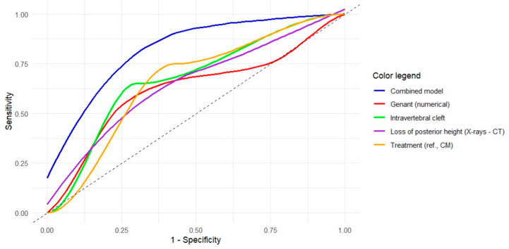
Objectives: We aimed to analyze potential predictors for the development of metachronous fractures (MFs) after osteoporotic vertebral fractures (OVFs), with particular focus on radiological variables obtained at initial X-rays and computed tomography (CT) examinations, treatment applied (conservative management [CM] versus percutaneous vertebroplasty [PV]), and fractures located at the thoracolumbar junction (T11-L2). Methods: We conducted a two-center, observational retrospective study, including patients with single-level OVFs treated with CM or VP. We collected socio-demographic, radiological and treatment-related variables. We performed descriptive and contrastive bivariate analyses based on the presence of MFs and univariate and multivariate logistic regression analyses to obtain adjusted and crude odds ratios (aOR and cOR, respectively) for predicting MFs. Finally, we performed receiver-operating characteristic (ROC) curve analyses to determine the discriminative power of the models obtained. Results: Of the 90 patients included, 20 (22.2%) developed one or more MFs (15 in CM and 5 in PV groups, respectively; p = 0.037). The treatment group (aOR for PV, 0.087; 95%CI, 0.015-0.379), presence of intravertebral cleft (aOR, 5.62; 95%CI, 1.84-19.2) and difference in posterior height loss between X-rays and CT (aOR, 0.926; 95%CI, 0.856-0.992) were identified as significant predictors for MFs, while Genant's numerical classification showed a trend toward significance (aOR, 1.97; 95%CI, 0.983-4.19; p = 0.064). A multivariate model combining these four variables showed optimal fitting and correctly discriminated over 80% of cases (AUC, 0.828; 95%CI, 0.725-0.930). Factors associated with MFs in thoracolumbar junction OVFs were intravertebral cleft, CM, posterior height loss in CT, and DGOU OF3 fractures. Conclusions: The presence of intravertebral cleft, a difference in posterior height loss between X-rays and CT equal to or lower than 2.4%, higher grades of Genant's numerical classification, and application of CM instead of PV are predictors of MFs. These findings improve our understanding of the factors involved in the development of MFs, but they need to be validated prospectively.

Keywords: conservative management; fracture; osteoporosis; percutaneous vertebroplasty; risk factor; spine.


Conflict of interest statement

The first and senior authors host a special issue in Diagnostics. They were not involved in the review process or editorial decisions related to the manuscript.

Figures

Figure 1
Figure 1
Examples of radiological variables assessed in our sample, from a 58-year-old woman (AC) and an 88-year-old man (D). Patient 1. X-rays ((A), magnified in (B)) and CT at diagnosis (C). The height of each wall (anterior, middle and posterior) was measured through lines parallel to the anterior vertebral walls. Relative measurements were obtained for each fractured vertebral body by dividing them between the mean of the respective measurements in the cephalad and caudal vertebral bodies. Note that some cases involved subtle findings such as the asymmetric, focal endplate depression depicted with a white arrow in (B). Patient 2. CT at diagnosis (D) showing an obvious subchondral defect (orange arrow) consistent with a large intravertebral cleft.
Figure 2
Figure 2
Examples of CT density measurements in a fractured vertebral body (arrow) and adjacent healthy vertebrae. The dashed lines with arrows indicate the level of different measurements in the axial plane. The dashed circles represent the region of interest (ROI) measured in the central part of the vertebral body.
Figure 3
Figure 3
Receiver operating characteristic (ROC) curves of the univariate and multivariate models obtained for the prediction of metachronous vertebral fractures. The colored curves represent the ROC curves for each significant independent variable based on the univariate logistic regression results (see legend). The blue curve refers to the multivariate model with 4 variables. The gray diagonal line corresponds to the reference of a random classification (line of no discrimination).
Figure 4
Figure 4
Illustrative example of a patient without metachronous fractures in our sample. (A) Initial X-rays. (B) Initial CT (MPR set at 1 mm). (C) Initial CT (MPR set at 100 mm for easier comparison with measurements on X-rays). (D) Follow-up X-rays. This is a 77-year-old woman with a biconcave, grade 1 (Genant’s classification), OF2 (DGOU’s classification) fracture of L1. The loss of posterior vertebral height (PVH) on CT was negative and the difference in loss of PVH was close to 0 (0.1%). She was treated with vertebroplasty (note a slight cement leak at T12-L1 intervertebral disc) and developed no metachronous fractures. In addition, follow-up X-rays 3 years later showed low-to-moderate local kyphosis (monosegmental Cobb angle of 15°) with preservation of sagittal balance (orange vertical line in (D)).
Figure 5
Figure 5
Illustrative example of a patient with metachronous fracture in our sample. (A) Initial X-rays. (B) Initial CT (MPR set at 1 mm). (C) Initial CT (MPR set at 100 mm for easier comparison with measurements on X-rays). (D) Follow-up X-rays. This is a 74-year-old woman with a biconcave, grade 2 (Genant’s classification), OF2 (DGOU’s classification) involving L1. The loss of posterior vertebral height (PVH) on CT was 9.5% and the difference in loss of PVH between X-rays and CT was 2%. She was managed conservatively and developed vertebral collapse and hyperkyphosis (monosegmental Cobb angle close to 30°) with loss of sagittal balance (sagittal vertical axis shown as an orange vertical line at the left panel in (D)). At 2-year follow-up, she developed L1 collapse and a metachronous fracture of T11 (yellow arrow in (D)).

Similar articles

References

    1. Xie L., Zhao Z.G., Zhang S.J., Hu Y. Bin Percutaneous Vertebroplasty versus Conservative Treatment for Osteoporotic Vertebral Compression Fractures: An Updated Meta-Analysis of Prospective Randomized Controlled Trials. Int. J. Surg. 2017;47:25–32. doi: 10.1016/j.ijsu.2017.09.021. - DOI - PubMed
    1. Yamauchi K., Adachi A., Kameyama M., Murakami M., Sato Y., Kato C., Kato T. A Risk Factor Associated with Subsequent New Vertebral Compression Fracture after Conservative Therapy for Patients with Vertebral Compression Fracture: A Retrospective Observational Study. Arch. Osteoporos. 2020;15:1–12. doi: 10.1007/s11657-019-0679-x. - DOI - PubMed
    1. Kelly M.A., McCabe E., Bergin D., Kearns S.R., McCabe J.P., Armstrong C., Heaney F., Carey J.J. Osteoporotic Vertebral Fractures Are Common in Hip Fracture Patients and Are Under-Recognized. J. Clin. Densitom. 2021;24:183–189. doi: 10.1016/j.jocd.2020.05.007. - DOI - PubMed
    1. Wang F., Sun R., Zhang S.D., Wu X.T. Comparison of Acute Single versus Multiple Osteoporotic Vertebral Compression Fractures in Radiographic Characteristic and Bone Fragility. J. Orthop. Surg. Res. 2023;18:1–10. doi: 10.1186/s13018-023-03874-7. - DOI - PMC - PubMed
    1. Kano S., Tanikawa H., Mogami Y., Shibata S.I., Takanashi S., Oji Y., Aoki T., Oba H., Ikegami S., Takahashi J. Comparison between Continuous and Discontinuous Multiple Vertebral Compression Fractures. Eur. Spine J. 2012;21:1867–1872. doi: 10.1007/s00586-012-2210-6. - DOI - PMC - PubMed

LinkOut - more resources