

Comparison of Resampling Methods and Radiomic Machine Learning Classifiers for Predicting Bone Quality Using Dual-Energy X-Ray Absorptiometry
- PMID: 39857059
- PMCID: PMC11763683
- DOI: 10.3390/diagnostics15020175
Comparison of Resampling Methods and Radiomic Machine Learning Classifiers for Predicting Bone Quality Using Dual-Energy X-Ray Absorptiometry
Abstract
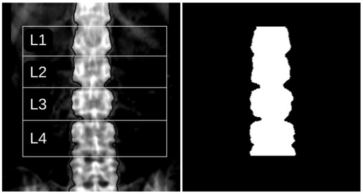
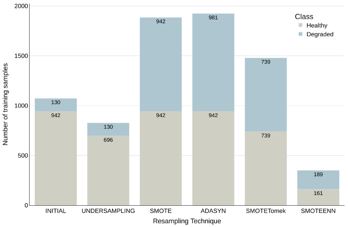
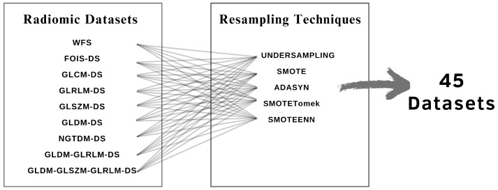
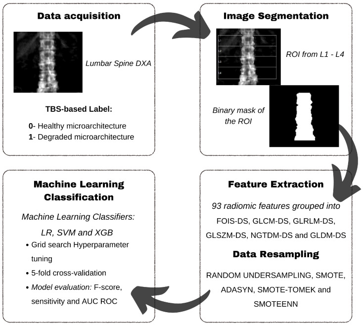
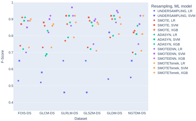
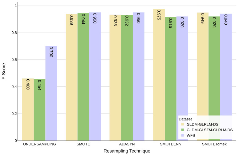
Background/Objectives: This study presents a novel approach, based on a combination of radiomic feature extraction, data resampling techniques, and machine learning algorithms, for the detection of degraded bone structures in Dual X-ray Absorptiometry (DXA) images. This comprehensive approach, which addresses the critical aspects of the problem, distinguishes this work from previous studies, improving the performance achieved by the most similar studies. The primary aim is to provide clinicians with an accessible tool for quality bone assessment, which is currently limited. Methods: A dataset of 1531 spine DXA images was automatically segmented and labelled based on Trabecular Bone Score (TBS) values. Radiomic features were extracted using Pyradiomics, and various resampling techniques were employed to address class imbalance. Three machine learning classifiers (Logistic Regression, Support Vector Machine (SVM), and XGBoost) were trained and evaluated using standard performance metrics. Results: The SVM classifier outperformed the other classifiers. The highest F-score of 97.5% was achieved using the Grey Level Dependence Matrix and Grey Level Run Length Matrix feature combination with SMOTEENN resampling, which proved to be the most effective resampling technique, while the undersampling method yielded the lowest performance. Conclusions: This research demonstrates the potential of radiomic texture features, resampling techniques, and machine learning methods for classifying DXA images into healthy or degraded bone structures, which potentially leads to improved clinical diagnosis and treatment.
Keywords: data resampling; dual energy X-ray absorptiometry; machine learning; radiomics; trabecular bone score.
Conflict of interest statement
The authors declare no conflicts of interest.
Figures
Similar articles
-
Application of machine learning model to predict osteoporosis based on abdominal computed tomography images of the psoas muscle: a retrospective study.BMC Geriatr. 2022 Oct 13;22(1):796. doi: 10.1186/s12877-022-03502-9. BMC Geriatr. 2022. PMID: 36229793 Free PMC article.
-
Diffusion-weighted MRI radiomics of spine bone tumors: feature stability and machine learning-based classification performance.Radiol Med. 2022 May;127(5):518-525. doi: 10.1007/s11547-022-01468-7. Epub 2022 Mar 23. Radiol Med. 2022. PMID: 35320464 Free PMC article.
-
Learning from Imbalanced Data: Integration of Advanced Resampling Techniques and Machine Learning Models for Enhanced Cancer Diagnosis and Prognosis.Cancers (Basel). 2024 Oct 8;16(19):3417. doi: 10.3390/cancers16193417. Cancers (Basel). 2024. PMID: 39410036 Free PMC article.
-
Comparative Studies on Resampling Techniques in Machine Learning and Deep Learning Models for Drug-Target Interaction Prediction.Molecules. 2023 Feb 9;28(4):1663. doi: 10.3390/molecules28041663. Molecules. 2023. PMID: 36838652 Free PMC article. Review.
-
COVID-19 diagnosis: A comprehensive review of pre-trained deep learning models based on feature extraction algorithm.Results Eng. 2023 Jun;18:101020. doi: 10.1016/j.rineng.2023.101020. Epub 2023 Mar 16. Results Eng. 2023. PMID: 36945336 Free PMC article. Review.
References
-
- Karunanithi R., Ganesan S., Panicker T., Korath M.P., Jagadeesan K. Assessment of bone mineral density by DXA and the trabecular microarchitecture of the calcaneum by texture analysis in pre-and postmenopausal women in the evaluation of osteoporosis. J. Med. Phys. 2007;32:161–168. doi: 10.4103/0971-6203.37481. - DOI - PMC - PubMed
-
- Sapthagirivasan V., Anburajan M., Mahadevan V. Bone trabecular analysis of femur radiographs for the assessment of osteoporosis using DWT and DXA. Int. J. Comput. Theory Eng. 2013;5:616. doi: 10.7763/IJCTE.2013.V5.760. - DOI
LinkOut - more resources
Full Text Sources
