. 2024 Dec 31;14(1):39.
doi: 10.3390/antiox14010039.

Nicotinamide Mononucleotide Restores NAD+ Levels to Alleviate LPS-Induced Inflammation via the TLR4/NF-κB/MAPK Signaling Pathway in Mice Granulosa Cells

Affiliations

Nicotinamide Mononucleotide Restores NAD+ Levels to Alleviate LPS-Induced Inflammation via the TLR4/NF-κB/MAPK Signaling Pathway in Mice Granulosa Cells

Mehboob Ahmed et al. Antioxidants (Basel). .

Abstract

Inflammation disrupts the normal function of granulosa cells (GCs), which leads to ovarian dysfunction and fertility decline. Inflammatory conditions such as polycystic ovary syndrome (PCOS), primary ovarian insufficiency (POI), endometriosis, and age-related ovarian decline are often associated with chronic low-grade inflammation. Nicotinamide mononucleotide (NMN) is an important precursor of NAD+ and has gained attention for its potential to modulate cellular metabolism, redox homeostasis, and mitigate inflammation. This study investigated the protective roles of NMN against lipopolysaccharide LPS-mediated inflammation in GCs. The results of this experiment demonstrated that LPS had negative effects on GCs in term of reduced viability and proliferation rates and upregulated the production of pro-inflammatory cytokines, including interleukin-1 beta (IL-1β), interleukin-6 (IL-6), cyclooxygenase-2 (Cox-2), and tumor necrosis factor-alpha (TNF-α). Notably, the levels of NAD+ and NAD+/NADH ratio in GCs were reduced in response to inflammation. On the other hand, NMN supplementation restored the NAD+ levels and the NAD+/NADH ratio in GCs and significantly reduced the expression of pro-inflammatory markers at both mRNA and protein levels. It also enhanced cell viability and proliferation rates of GCs. Furthermore, NMN also reduced apoptosis rates in GCs by downregulating pro-apoptotic markers, including Caspase-3, Caspase-9, and Bax while upregulating anti-apoptotic marker Bcl-2. NMN supplementation significantly reduced reactive oxygen species ROS and improved steroidogenesis activity by restoring the estradiol (E2) and progesterone (P4) levels in LPS-treated GCs. Mechanistically, this study found that NMN suppressed the activation of the TLR4/NF-κB/MAPK signaling pathways in GCs, which regulates inflammatory processes. In conclusion, the findings of this study revealed that NMN has the potential to reduce LPS-mediated inflammatory changes in GCs by modulating NAD+ metabolism and inflammatory signaling pathways. NMN supplementation can be used as a potential therapeutic agent for ovarian inflammation and related fertility disorders.

Keywords: NAD+; NMN; ROS; apoptosis; granulosa cell; inflammation; steroidogenesis.


Conflict of interest statement

The authors declare no conflicts of interest.

Figures

Figure 1
Figure 1
Effects of LPS on granulosa cells viability, proliferation, mRNA expression, and protein levels of pro-inflammatory markers. (A) Percentage of granulosa cell (GC) viability following treatment with varying concentrations of LPS (0.1, 1, 5, 10, 20, and 100 µg/mL) in culture media, showing a significant reduction at concentrations ≥ 10 µg/mL. (B) GC proliferation presented as the number of cells, with a significant decrease observed at 10 µg/mL LPS and higher. (C) mRNA expression of TLR4, IL-1β, IL-6, Cox-2, and TNF-α analyzed by qPCR, demonstrating significant upregulation in LPS-treated GCs compared to the control. (D) Western blot representative images and analysis showing increased protein expression of TLR4, phosphorylated NF-κB (p-NF-κB), COX-2, and TNF-α in response to LPS treatment, while GAPDH serves as the loading control. Values are expressed as mean ± SEM. Compared to the control group, * p < 0.05, ** p < 0.01, and *** p < 0.001.
Figure 2
Figure 2
Effects of LPS on the NAD+ levels, NADH levels, and NAD+/NADH ratio in granulosa cells. (A) The NAD+ and NADH levels were measured in GCs (~1 × 10⁶ cells). (A) NAD+ levels in GCs after LPS treatment, showing a significant decrease compared to the control. (B) The NADH levels remained unchanged between the control and LPS-treated groups, indicating no significant effect of LPS on the NADH level. (C) The NAD+/NADH ratio in GCs, demonstrating a significant reduction in LPS-treated cells compared to the control. These data illustrate the metabolic shift induced by LPS in GCs, primarily driven by a reduction in the NAD+ levels and the NAD+/NADH ratio. Values are expressed as mean ± SEM. Compared to the control group, ** p < 0.01, and *** p < 0.001.
Figure 3
Figure 3
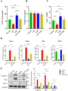
Effects of NMN supplementation on LPS-treated granulosa cells viability, proliferation, mRNA expression, and protein levels of pro-inflammatory markers. (A) Percentage of cell viability following treatment with varying concentrations of NMN (1, 10, 100, 500, 1000, and 2000 µM) in LPS-treated GCs, showing a significant increase at 100 µM, with the highest viability observed at 500 and 1000 µM. (B) GC proliferation presented as the number of cells, with a significant improvement observed at 100 µM and the highest rates observed at 500 and 1000 µM of NMN in LPS-treated GCs. (C) mRNA expression of IL-1β, IL-6, Cox-2, and TNF-α analyzed by qPCR, showing significant upregulation in LPS-treated GCs compared to the control, and a marked reduction in the LPS + NMN group as compared to LPS-treated GCs (D) Western blot representative images and analysis showing a reduced protein expression of Cox-2 and TNF-α in LPS + NMN-treated GCs compared to the LPS group. GAPDH was used as the loading control. Values are expressed as mean ± SEM. ns = non-significant; p ≥ 0.05, * p < 0.05, ** p < 0.01, and *** p < 0.001.
Figure 4
Figure 4
Effects of NMN supplementation on the NAD+ levels, NADH levels, and NAD+/NADH ratio in granulosa cells. The NAD+ and NADH levels were measured in GCs (~1 × 10⁶ cells) across four groups: Control, LPS, NMN, and LPS + NMN. (A) The NAD+ levels significantly decreased in LPS-treated GCs compared to the controls, while NMN supplementation restored the NAD+ levels to baseline. (B) The NADH levels showed no significant differences across the experimental groups. (C) The NAD+/NADH ratio, reduced by LPS treatment, was significantly restored with NMN supplementation. (D) The mRNA expression levels of apoptosis-related markers were assessed by qPCR. LPS treatment significantly upregulated the pro-apoptotic markers, including Caspase-3 (Cas-3), Caspase-9 (Cas-9), and Bax, while reducing the expression of the anti-apoptotic marker Bcl-2 compared to the control group. NMN supplementation (LPS + NMN) significantly reduced the expression of Cas-3, Cas-9, and Bax while restoring Bcl-2 expression in LPS-treated GCs. (E) Western blot analysis confirmed the protein-level changes in the apoptosis markers. LPS treatment increased the protein expression of Caspase-3 and Bax and reduced Bcl-2 expression compared to the control group. NMN supplementation significantly decreased the Caspase-3 and Bax levels and increased Bcl-2 protein expression in LPS-treated GCs. Data are presented as mean ± SEM with significance levels indicated: ns = non-significant; p ≥ 0.05, * p < 0.05, ** p < 0.01, and *** p < 0.001.
Figure 5
Figure 5
Effects of NMN supplementation on the ROS levels and steroidogenesis activity in granulosa cells. (A) Intracellular ROS levels were measured in GCs to assess oxidative stress. LPS-treated GCs showed a significant increase in ROS compared to the control group, while NMN supplementation reduced ROS in the LPS + NMN group, indicating protective effects against oxidative stress. (B,C) The levels of estradiol (E2) and progesterone (P4) in GCs were also evaluated. LPS treatment significantly reduced E2 and P4 production, whereas NMN supplementation restored their levels to the control values, supporting steroidogenic function. Values are presented as mean ± SEM, with significance levels indicated: ns = non-significant; p ≥ 0.05, * p < 0.05, ** p < 0.01, and *** p < 0.001.
Figure 6
Figure 6
Effects of NMN on TLR4/NF-κB p65/MAPK pathway activation in granulosa cells. Western blot analysis shows that LPS treatment increased the expression of TLR4 (relative to GAPDH), p-NF-κB p65 (relative to NF-κB p65), p-ERK1/2 (relative to ERK1/2), p-JNK (relative to JNK), and p-P38 (relative to GAPDH) compared to the control group, while NMN supplementation in LPS-treated GCs reduced the expression of these proteins, indicating that NMN alleviates LPS-induced inflammatory signaling through the TLR4/NF-κB p65/MAPK pathway. Values are expressed as mean ± SEM; with significance levels indicated: ns = non-significant; p ≥ 0.05, * p < 0.05, ** p < 0.01, and *** p < 0.001.
Figure 7
Figure 7
Graphical illustration of TLR4/NF-κB p65/MAPK pathway activation and inhibition by NMN. This illustration depicts the activation of the TLR4/NF-κB p65 and MAPK pathways in granulosa cells in response to LPS. LPS binding to TLR4 triggers downstream signaling, leading to NF-κB p65 phosphorylation and MAPK pathway activation, as indicated by the increased p-ERK1/2, p-JNK, and p-P38. The ROS levels and apoptosis are also increased in response to LPS-mediated TLR4 activation. NMN supplementation counteracts this activation by increasing the NAD+ levels and reducing TLR4 signaling and subsequently downregulating NF-κB p65 and MAPK phosphorylation, effectively mitigating the inflammatory responses, ROS production, and apoptosis markers. This graphical overview highlights the anti-inflammatory and anti-apoptotic roles of NMN and NAD+ in cellular signaling.

References

    1. Wang Y., Yang Q., Wang H., Zhu J., Cong L., Li H., Sun Y. NAD+ deficiency and mitochondrial dysfunction in granulosa cells of women with polycystic ovary syndrome. Biol. Reprod. 2021;105:371–380. doi: 10.1093/biolre/ioab078. - DOI - PubMed
    1. Witchel S.F., Oberfield S.E., Peña A.S. Polycystic ovary syndrome: Pathophysiology, presentation, and treatment with emphasis on adolescent girls. J. Endocr. Soc. 2019;3:1545–1573. doi: 10.1210/js.2019-00078. - DOI - PMC - PubMed
    1. Cattelan A., Ceolotto G., Bova S., Albiero M., Kuppusamy M., De Martin S., Semplicini A., Fadini G.P., de Kreutzenberg S.V., Avogaro A. NAD+-dependent SIRT1 deactivation has a key role on ischemia–reperfusion-induced apoptosis. Vasc. Pharmacol. 2015;70:35–44. doi: 10.1016/j.vph.2015.02.004. - DOI - PubMed
    1. Kang H., Park Y.-K., Lee J.-Y. Nicotinamide riboside, an NAD+ precursor, attenuates inflammation and oxidative stress by activating sirtuin 1 in alcohol-stimulated macrophages. Lab. Investig. 2021;101:1225–1237. doi: 10.1038/s41374-021-00599-1. - DOI - PubMed
    1. Zhao Y., Zhang J., Zheng Y., Zhang Y., Zhang X.J., Wang H., Du Y., Guan J., Wang X., Fu J. NAD+ improves cognitive function and reduces neuroinflammation by ameliorating mitochondrial damage and decreasing ROS production in chronic cerebral hypoperfusion models through Sirt1/PGC-1α pathway. J. Neuroinflamm. 2021;18:207. doi: 10.1186/s12974-021-02250-8. - DOI - PMC - PubMed

LinkOut - more resources