Review
. 2025 Jan 8;17(2):181.
doi: 10.3390/cancers17020181.

Very High-Energy Electron Therapy Toward Clinical Implementation

Affiliations
Review

Very High-Energy Electron Therapy Toward Clinical Implementation

Costanza Maria Vittoria Panaino et al. Cancers (Basel). .

Abstract

The use of very high energy electron (VHEE) beams, with energies between 50 and 400 MeV, has drawn considerable interest in radiotherapy due to their deep tissue penetration, sharp beam edges, and low sensitivity to tissue density. VHEE beams can be precisely steered with magnetic components, positioning VHEE therapy as a cost-effective option between photon and proton therapies. However, the clinical implementation of VHEE therapy (VHEET) requires advances in several areas: developing compact, stable, and efficient accelerators; creating sophisticated treatment planning software; and establishing clinically validated protocols. In addition, the perspective of VHEE to access ultra-high dose-rate regime presents a promising avenue for the practical integration of FLASH radiotherapy of deep tumors and metastases with VHEET (FLASH-VHEET), enhancing normal tissue sparing while maintaining the inherent dosimetric advantages of VHEET. However, FLASH-VHEET systems require validation of time-dependent dose parameters, thus introducing additional technological challenges. Here, we discuss recent progress in VHEET research, focusing on both conventional and FLASH modalities, and covering key aspects including dosimetric properties, radioprotection, accelerator technology, beam focusing, radiobiological effects, and clinical outcomes. Furthermore, we comprehensively analyze initial VHEET in silico studies on coverage across various tumor sites.

Keywords: FLASH radiotherapy; VHEE; external beam radiotherapy.


Conflict of interest statement

The authors declare no conflicts of interest.

Figures
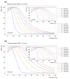
Figure 2
Figure 2
Longitudinal on-axis (main) and integral (insert) dose distributions of collimated beams impinging on a water phantom (with vacuum beforehand). The beam energy varies from 50 to 300 MeV in 50 MeV increments. The beam diameters are (a) 1 cm and (b) 10 cm.
Figure 1
Figure 1
(a) Longitudinal on-axis dose distribution of a collimated VHEE parallel beam impinging on a water phantom (with vacuum beforehand); the beam has an energy of 200 MeV and a 1 cm diameter, showing the dmax (maximum dose depth), PFO (proximal fall-off at 90% of the dose at dmax), and TR (therapeutic range at 90% of the dose at dmax). The transversal dose distribution is shown at depths of (b) dmax, (c) 20, and (d) 30 cm. The LP (lateral penumbra) is the distance between the 90% and 20% intensity levels, whereas the BW (beam width) is the width at 90% of the maximum dose value at that depth.
Figure 3
Figure 3
FLASH vs. CONV-RT modality: inter-dependent temporal parameters characterizing the entire RT treatment, a single fraction, and a single pulse.
Figure 4
Figure 4
Facilities currently being used worldwide for VHEE beam production are categorized as follows: Green for RF-based research linear accelerators (RF linacs) [12,18,19,24,31,62,64,66,67,68,69,70,71,72,73,74], yellow for compact FLASH VHEET-dedicated RF linac platforms [10,11], and red for research laser–plasma accelerators exploiting the Wakefield effect [13,15,16,21,31,63,75,76,77,78,79]. Listed under each facility are the papers based on research conducted at that center.
Figure 5
Figure 5
Schematic representation of a linear accelerator (linac) and its main components: Electrons are generated by a particle source within a vacuum chamber, using either a cold cathode, a hot cathode, a photocathode, or an RF source. A microwave source, such as a klystron or magnetron, powers the accelerating waveguide, a series of open-ended cylindrical electrodes guiding the electron beam toward the diagnostic and manipulation (focusing) area. The arrows represent the electric field oscillating (red/blue) direction between the electrodes of the linac powered by the RF generator. Finally, the VHEE beam reaches the user beamline.
Figure 6
Figure 6
Schematic representation of a laser Wakefield accelerator with its main components. An ultra-short (∼10–40 fs) laser pulse is generated, amplified, and transported to the interaction area. The laser pulse is then focused on a gas target, usually by an off-axis parabolic mirror. Electrons are accelerated within the plasma and propagate in the forward direction.
Figure 7
Figure 7
Particle-in-cell simulation [102] artwork of laser Wakefield electron acceleration. The laser pulse propagates in an underdense “transparent” plasma, generating a strong longitudinal electric field, whose velocity is close to c, the Wakefield. When electrons are trapped within the accelerating region of the electric field, their energy is boosted to very high values over very short distances.
Figure 8
Figure 8
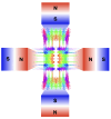
Quadruple magnetic field lines. Colors represent the magnetic field direction and strength.
Figure 9
Figure 9
VHEE beam focusing: (a) Focusing and (b) defocusing magnetic quadrupoles, graphic rendering from the thin lens approximation, and matrix formalism. (c) Drift graphic rendering and matrix formalism. The final focusing can be symmetric (d), meaning the beam converges along both planes or asymmetrically (e), with the beam converging along one plane only.

References

    1. Hogstrom K.R., Almond P.R. Review of electron beam therapy physics. Phys. Med. Biol. 2006;51:R455. doi: 10.1088/0031-9155/51/13/R25. - DOI - PubMed
    1. Gahbauer R., Landberg T., Chavaudra J., Dobbs J., Gupta N., Hanks G., Horiot J.-C., Johansson K.-A., Möller T., Naudy S., et al. Prescribing, Recording, and Reporting Electron Beam Therapy. J. ICRU. 2004;4:5–9. doi: 10.1093/jicru/ndh001. - DOI
    1. DesRosiers C., Moskvin V., Bielajew A.F., Papiez L. 150-250 MeV electron beams in radiation therapy. Phys. Med. Biol. 2000;45:1781. doi: 10.1088/0031-9155/45/7/306. - DOI - PubMed
    1. Yeboah C., Sandison G.A., Moskvin V. Optimization of intensity-modulated very high energy (50–250 MeV) electron therapy. Phys. Med. Biol. 2002;47:1285. doi: 10.1088/0031-9155/47/8/305. - DOI - PubMed
    1. Yeboah C., Sandison G.A. Optimized treatment planning for prostate cancer comparing IMPT, VHEET and 15 MV IMXT. Phys. Med. Biol. 2002;47:2247. doi: 10.1088/0031-9155/47/13/305. - DOI - PubMed

LinkOut - more resources