. 2025 Jan 20;15(2):275.
doi: 10.3390/ani15020275.

The Sublingua of Lemur catta and Varecia variegata: Only a Cleaning Function?

Affiliations

The Sublingua of Lemur catta and Varecia variegata: Only a Cleaning Function?

Juan Francisco Pastor et al. Animals (Basel). .

Abstract

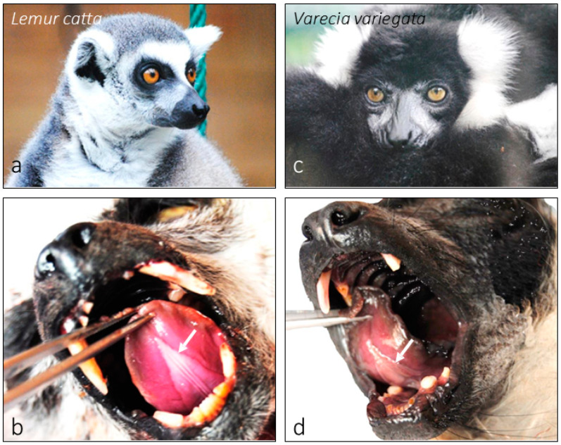
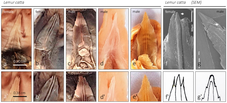
The sublingua is an anatomical structure located under the tongue. This rare organ can be present in some animals as a rudimentary structure, but among prosimian primates, such as lemurs and lorises, it is fully developed. In addition to the sublingua, prosimians have modified lower incisors and canines called "dental comb". The anatomy of sublingua has been studied macro and microanatomically since the early 19th century. Most authors argue that the sublingua is an oral morphological adaptation to develop a toothbrush's role in cleaning the dental comb. However, others assert that the functional role has yet to be established. Comparative studies of macro and microanatomy are scarce or incomplete for primates; thus, the putative function remains unclear. To better understand the functional significance of the sublingua, we studied this structure in Lemur catta and Varecia variegata specimens using histochemical staining techniques and scanning electron microscopy with microanalysis. The new data obtained provide a fuller picture of the role assigned to sublingua so far, which could be more complex. In light of the morphological findings, we should consider additional roles/functions of the sublingua, including but not limited to food processing, grooming or social behavior.

Keywords: Lemur catta; Varecia variegata; lemur; scanning electron microscopy; sublingua.


Conflict of interest statement

The authors declare that they have no known competing financial interests or personal relationships that could have appeared to influence the work reported in this paper.

Figures

Figure 1
Figure 1
Images of Lemur catta (a,b) and Varecia variegata (c,d). Images (a,c) were taken from specimens hosted in the Santillana del Mar Zoo. Images (b,d) show the position of the sublingua in the oral cavity under the tongue.
Figure 2
Figure 2
The ventral aspect of the unfixed tongue and sublingua of Lemur catta (a,b) and Varecia variegata (c,d) shows the comparative size of such structures. The surface of the Varecia variegata sublingua presents several transversal folds similar to the folds that were previously described on the tongue’s dorsal surface by Pastor et al. [3], which was related to the way of feeding.
Figure 3
Figure 3
The images (Lemur catta on the left and Varecia variegata on the right) display the lower jaw or mandibular bone with the so-called “dental comb” (modified lower incisors and canines), the surface against which the sublingua would act as a toothbrush according to several classical studies. The images placed to the right of the images identified with the specie and gender correspond to the dorsal view of the same sample.
Figure 4
Figure 4
Images of the internal structure of the sublingua of Lemur catta stained with Masson’s trichrome. On the left (a), we summarize the approximate plane of the transversal cross-section shown on the right (be).
Figure 5
Figure 5
Ventral aspect of the sublingua of several Lemur catta specimens ((ae) and slightly enlarged in the second row of images (a’e’)). The sublingua’s tip could be observed. SEM images of a female (f) and male (g) specimen magnify the morphology of the tip. The drawings presented in (f’) (female) and (g’) (male) summarize the differences found between genders (3 females versus 4 males) regarding the morphology of the tip. In females, the central rod reaches the most cranial part of the tip, which is not the case in males. Such a comparison cannot be made in Varecia variegata because one of the 2 female specimens studied has a damaged and incomplete tip.
Figure 6
Figure 6
SEM images of Lemur catta sublingua’s ventral surface. The tip of a male specimen with some areas magnified (af). In the “(i)” low magnification SEM image, we could appreciate an anterior smooth surface (left side) and a posterior rough surface (right side). Images (g,h) show the ventral smooth anterior surface with a keratinized area with some porous zones (magnified in (h)) presented sparsely along such surfaces. With different magnifications (images (jl)), the morphology of the rough posterior surface caused by several pseudo papillae, similar to mechanical ones, could be appreciated. Scale bar image (h), 1 μm.
Figure 7
Figure 7
Analysis of element composition based on backscattered electrons with the point of measurement (spectrum) in Lemur catta samples. We focused on keratinized areas with and without porous surfaces, described in Figure 6g,h. On keratinized and nonporous surfaces, the analysis yielded the presence of carbon and oxygen (not shown in the figure). In contrast, we also found iron in the composition of the substance in the porous surface (image (b)). On the non-porous keratinized surface, we found a crystallized substance, which through analysis was determined to be silica in composition (image (a)), making it compatible with the grains of sand found in the animal’s habitat. Toluidine blue-stained transverse sections focused on the keratinized layer of Lemur catta sublingua with non-keratinized zones (c,d).
Figure 8
Figure 8
Images of the internal structure of the sublingua of Varecia variegata stained with Masson’s trichrome. On the left (a), we summarize the approximate plane of the transversal cross-section shown on the right (be). Note that the inner structure is filled with cavities (be).
Figure 9
Figure 9
Different images were obtained using the SEM technique from the ventral surface of the sublingua in Varecia variegata. In image (b), we displayed the zone studied. We found bright (a,d,f) and dark (c,e,g) areas. We could observe a porous surface in bright areas without any substance filling them. In contrast, we show that the porous surface is filled with a substance in the dark areas. The substance was analyzed, and the results are displayed in Figure 9.
Figure 10
Figure 10
Analysis of element composition based on the backscattered electrons technique and the point of measurement (spectrum) in Varecia variegata sublingua samples, focusing on the bright and dark areas described in Figure 9. The analysis of a porous region without any filling substance yielded only carbon and oxygen (a), whereas the porous region filled with a substance also yielded chlorine in the composition (b). Masson’s trichrome-stained sections of a Varecia variegata sublingua transverse section show cavernous-like structures with projections towards the keratinized layer (c,d).

References

    1. Rommel C. Sublingual structures in primates. Part 1: Prosimiae, Platyrrhini and Cercopithecinae. Gegenbaurs Morphol. Jahrb. 1981;127:153–1752. (In German) - PubMed
    1. Curvier F.G. Histoire Naturelle des Mammiferes. Chez à. Belin (Libraire-Éditeur); Paris, France: 1829. p. 218.
    1. Pastor J.F., Muchlinski M.N., Potau J.M., Casado A., García-Mesa Y., Vega J.A., Cabo R. The Tongue in Three Species of Lemurs: Flower and Nectar Feeding Adaptations. Animals. 2021;11:2811. doi: 10.3390/ani11102811. - DOI - PMC - PubMed
    1. Hofer H.O., Castenholz A., Zöltzer H. The sublingua and tongue of Tupaia (Scandentia, mammalia): A scanning electron microscopy study. Folia Primatol. 1993;60:185–194. doi: 10.1159/000156692. - DOI - PubMed
    1. Pocock R.I. On the External Characters of the Lemurs and of Tarsius. Proc. Zool. Soc. Lond. 1918;88:19–53. doi: 10.1111/j.1096-3642.1918.tb02076.x. - DOI

LinkOut - more resources