Review
. 2025 Jan 10;15(1):102.
doi: 10.3390/biom15010102.

Tetrahydrobiopterin in Myalgic Encephalomyelitis/Chronic Fatigue Syndrome: A Friend or Foe?

Affiliations
Review

Tetrahydrobiopterin in Myalgic Encephalomyelitis/Chronic Fatigue Syndrome: A Friend or Foe?

A F M Towheedur Rahman et al. Biomolecules. .

Abstract

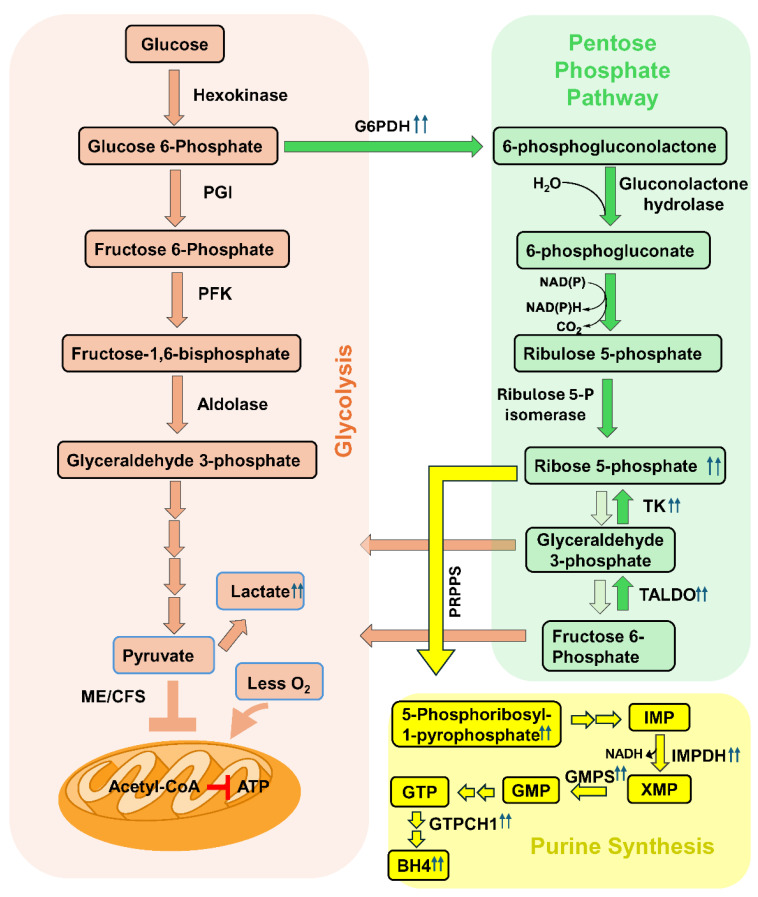
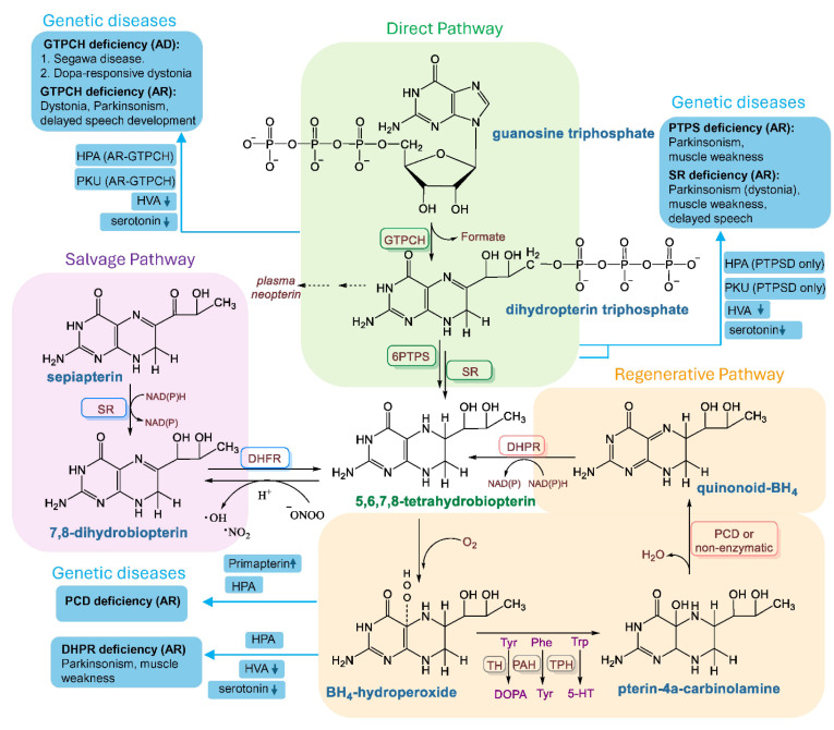
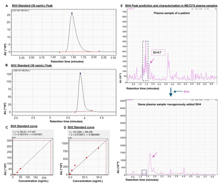
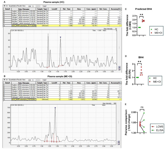
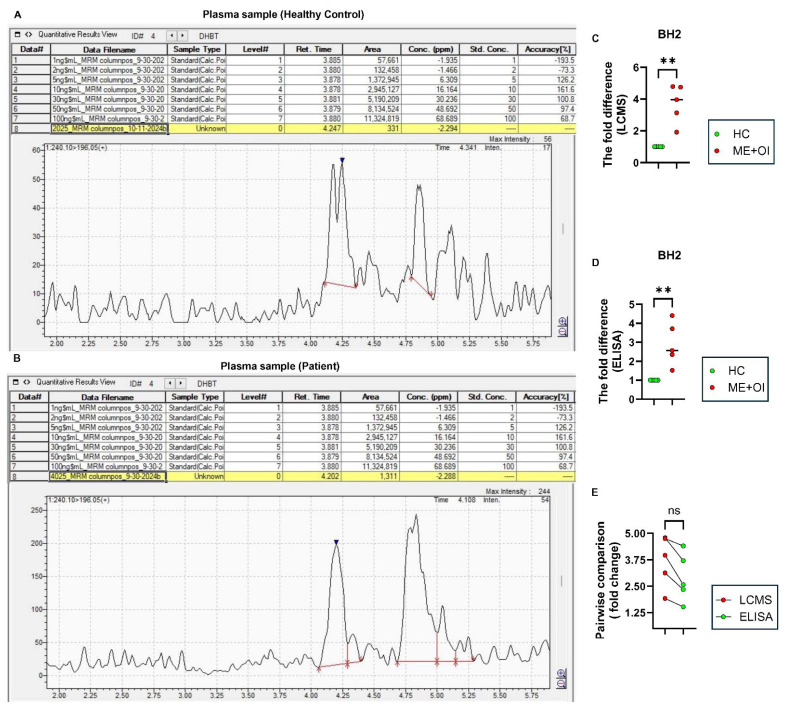
Myalgic Encephalomyelitis or Chronic Fatigue Syndrome (ME/CFS) is a chronic multisystem disease characterized by severe muscle fatigue, pain, dizziness, and brain fog. The two most common symptoms are post-exertional malaise (PEM) and orthostatic intolerance (OI). ME/CFS patients with OI (ME+OI) suffer from dizziness or faintness due to a sudden drop in blood pressure while maintaining an upright posture. Clinical research has demonstrated that patients with OI display severe cardiovascular abnormalities resulting in reduced effective blood flow in the cerebral blood vessels. However, despite intense investigation, it is not known why the effective cerebral blood flow is reduced in OI patients. Based on our recent findings, we observed that tetrahydrobiopterin (BH4) metabolism was highly dysregulated in ME+OI patients. In the current review article, we attempted to summarize our recent findings on BH4 metabolism to shed light on the molecular mechanisms of OI.

Keywords: ME/CFS; autophagy; dihydrobiopterin (BH2); orthostatic intolerance (OI); oxidative stress; pentose phosphate pathway (PPP); tetrahydrobiopterin (BH4).


Conflict of interest statement

A.R., S.B. and C.G.G. are employees of Simmaron Research INC, a 501C non-profit research organization. The authors declare no conflicts of interest.

Figures

Figure 5
Figure 5
Integrated metabolic pathways of BH4 metabolism in ME/CFS pathogenesis. (Red enclosure) The glycolysis pathway of glucose metabolism leads to the formation of pyruvate. Pyruvate is transported to mitochondria, converted to acetyl CoA, and metabolized by the TCA cycle, generating reduced intermediates for ATP production. The utilization of glucose by glycolysis is compromised in ME/CFS pathogenesis. Reduced oxygen consumption by mitochondria was reported to uncouple the electron transport chain for ATP synthesis. (Green enclosure) Augmented pentose phosphate pathway by the enzymic activation of glucose-6-phosphate dehydrogenase (G6PDH), Transketolase (TK), and transaldolase (TALDO). Reduced oxygen consumption by mitochondria and enhanced bioavailability of NADPH may induce the reduction potential in the cell, causing reversible enzymes TALDO and TK to increase the biogenesis of ribose-5-phosphate (R5P). (Yellow enclosure) Synthesis of purine metabolites. Phosphoribosyl pyrophosphate synthase (PRPPS) enzyme converts R5P to inosine-5-monophosphate (IMP), which is converted to GMP by the successive enzymic actions of IMP dehydrogenase (IMPDH) and GMP synthase (GMPS). GTP is synthesized by GTP, which generates BH4 via the direct pathway by the enzymic action of GTPCH1. R5P is the direct precursor for purine biosynthesis. Once the bioavailability of R5P increases and its utilization via glycolysis is inhibited, it is expected that the downstream formation of purine metabolites via the nucleotide biosynthetic pathway (yellow enclosure) will be operative. Phosphoribosyl pyrophosphate synthase (PRPPS) is the first enzyme of the purine biosynthetic pathway that catalyzes the synthesis of 5-phosphoribosyl-1-pyrophosphate (PRPP) from R5P. Previous studies reported that the induction of hypoxia enhanced the enzymic activity of PRPPS [52]. Accordingly, our biochemical assay also identified a very strong level of PRPP in the microglial cells when a hypoxic or less-oxygenated environment was implemented [21]. Moreover, we also demonstrated that inosine-5-monophosphate dehydrogenase (IMPDH), a rate-limiting enzyme of purine biosynthesis, and GMP synthase (GMPS), another key enzyme of purine biogenesis, were upregulated in hypoxic condition. Interestingly, inhibition of the non-oxidative PPP by taldo1 siRNA also attenuated the expression of both IMPDH and GMPS in microglia. Collectively, these observations demonstrated that the augmentation of the non-oxidative PPP by hypoxia-induced strong reductive potential guided R5P for purine biogenesis.
Figure 1
Figure 1
Biosynthesis of BH4 and genetic diseases due to BH4 deficiency. (Green enclosure) De novo or direct pathway of BH4 biosynthesis. Guanosine triphosphate (GTP) is enzymatically converted to BH4 by the successive enzymic actions of GTP cyclohydrolase (GTPCH1), 6-pyruvoyl-tetrahydrobiopterin synthase (6PTPS or PTPS), and sepiapterin reductase (SR). Autosomal dominant (AD) and autosomal recessive (AR) mutations of the gtpch1 gene cause BH4 deficiency via the de novo biogenesis pathway. PTPS deficiency and SR mutations are two other AR traits for BH4 deficiency. HPA = hyperphenylalaninemia, PKU = phenylketonuria, HVA = homovanillic acid. (Purple enclosure) Salvage pathway of BH4 biosynthesis. In this metabolic pathway, sepiapterin is first converted to 7,8-dihydrobiopterin (BH2) by sepiapterin reductase (SR) and then BH2 is converted to BH4 by dihydrofolate reductase (DHFR). BH4 is non-enzymically converted to BH2, contributing to the generation of reactive oxygen species. (Orange enclosure) Regenerative pathway. In the regenerative pathway, BH4 is regenerated in two reactions. BH4 is first oxidized to BH4-hydroperoxide and then converted to pterin-4a-carbinolamine during catalysis by aromatic amino acid hydroxylases such as tyrosine, phenylalanine, and tryptophan hydroxylases. Pterin-4a-carbinolamine is then converted to q-BH2 (Quinonoid BH2) by pterin-4a-carbinol- amine dehydratase (PCD) and then re-cycled back to BH4 by dihydropteridine reductase (DHPR). PCD deficiency (AR) causes the upregulation of the pterin metabolite primapterin and HPA. DHPR deficiency (AR) causes the loss of HVA, serotonin, and HPA. The flowchart of the BH4 metabolism is illustrated as described in Chapter 6 of the book “Nitric Oxide (2nd edition)” [23].
Figure 2
Figure 2
The potential challenge of detecting BH4 in plasma samples by a triple-quad LCMS-8040 strategy. (A) Representative peaks for purified BH4 (30 ng/mL; Cat# T4425-5MG; Millipore Sigma Aldrich, St. Louis, MO, USA) and (B) BH2 (30 ng/mL; Cat# 37272-10MG; Millipore Sigma Aldrich) dissolved in 0.002% formic acid in D.I. water. Ionization energy method = electrospray ionization; column type = Gemini 5 µm C18 110 Å LC Column 150 × 2 mm (Cat# 00F-4435-B0, Phenomenex, Torrance, MD, USA); mobile phase composition = binary solvent system, solvent A: 0.002% formic acid in water; solvent B: 0.002% formic acid in methanol. Standard curves for (C) BH4 and (D) BH2. (E) The electrospray ionization (nebulizing gas flow 2 L/min and drying gas flow 15 L/min) and heat (DL temperature 250 °C and heat block temperature 400 °C) generated during the run destabilized BH4. Therefore, it was extremely difficult to identify the BH4 peak. The demonstrated peak (pink arrow; upper spectrogram) is too small to be considered as a real peak. However, the exogenous addition of BH4 increased the peak height (pink arrow; lower spectrogram) without altering the adjacent peaks (enclosed in a dotted rectangle), suggesting that the peak could be a BH4 peak. In chromatograms, AU = arbitrary unit and “*104” of AU indicated 10,000 times of the displayed unit.
Figure 3
Figure 3
The comparative analysis of BH4 by LCMS and ELISA. Freshly harvested plasma samples were analyzed for LCMS and ELISA to compare the relative levels of BH4 (blue arrowheads) between healthy control (HC) and ME/CFS with orthostatic intolerance (ME+OI) groups (n = 5 per group). Representative mass spectrograms are displayed after analyzing plasma samples of (A) HC and (B) age-/gender-matched ME+OI subjects. The fold difference of BH4 levels was analyzed between the HC and age-/gender-matched ME+OI cases by (C) LCMS and (D) ELISA. The Mann–Whitney U test demonstrates the significance of the mean between the two groups with ** p < 0.01 = 0.0079. (E) Pairwise analyses of the same samples between LCMS and ELISA displays a similar trend (ns = no significance). In each table, the numbers under “Area” column are annotated by comma (,) for every 1000 unit. All other numbers under Retention time, concentrations, and accuracy columns are represented in decimal unit.
Figure 4
Figure 4
The comparative analysis of BH2 by LCMS and ELISA. Freshly harvested plasma samples were analyzed for LCMS and ELISA to compare the relative levels of BH4 (blue arrowheads) between healthy control (HC) and ME/CFS with orthostatic intolerance (ME+OI) groups (n = 5 per group). Representative mass spectrograms are displayed after analyzing plasma samples of (A) HC and (B) age-/gender-matched ME+OI subjects. The negative value in concentration (ppm) means that the BH2 peak area of the sample is below the chosen range of standard concentrations. It does not mean that BH2 was undetected. In these cases, area is considered. The fold difference of BH2 levels was analyzed between the HC and age-/gender-matched ME+OI cases by (C) LCMS and (D) ELISA. The Mann-Whitney U test demonstrates the significance of the mean between the two groups with ** p < 0.01 = 0.0079. (E) Pairwise analyses of the same samples between LCMS and ELISA displays a similar trend (ns = no significance). In each table, the numbers under “Area” column are annotated by comma (,) for every 1000 unit. All other numbers under Retention time, concentrations, and accuracy columns are represented in decimal unit.
Figure 6
Figure 6
Non-enzymic conversion of BH4 to BH2. A schema that displays a potential metabolic conversion of BH4 to BH2. Nucleophilic attack by peroxynitrite (OONO) at the N5 hydrogen of BH4 may generate a trihydrobiopterin intermediate, releasing OH and NO2 radicals. BH3 quickly converts to BH2, potentially by generating a hydride ion and further increasing the reduction potential in the cell.
Figure 7
Figure 7
The metabolic contributions of elevated biopterins in the pathogenesis of ME/CFS. Reduced mitochondria consumption of oxygen followed by the impairment of the electron transport chain increases electron pressure in mitochondria, affects the redox balance in cells, causing the augmentation of the non-oxidative pentose phosphate pathway, and may lead to the dysregulation of biopterin homeostasis. The potential redox imbalance increases reactive stress in the endoplasmic reticulum and disrupts the glutathione homeostasis, causing protein misfolding. Reactive stress also activates mTORC1, leading to autophagy impairment. Cellular increase in reductive potential causes the inactivation of NRF2 and downregulation of anti-oxidant genes in mitochondria. All these phenomena may lead to the augmentation of stress and inflammation.

References

    1. Kaufman S. Ciba Foundation Symposium 22-Aromatic Amino Acids in the Brain. Wiley Online Library; Hoboken, NJ, USA: 1974. Properties of the pterin-dependent aromatic amino acid hydroxylases; pp. 85–115.
    1. Tayeh M., Marletta M. Macrophage oxidation of L-arginine to nitric oxide, nitrite, and nitrate: Tetrahydrobiopterin is required as a cofactor. J. Biol. Chem. 1989;264:19654–19658. doi: 10.1016/S0021-9258(19)47163-9. - DOI - PubMed
    1. Hopkins F.G. Note on a yellow pigment in butterflies. Nature. 1889;40:335
    1. Bendall J.K., Douglas G., McNeill E., Channon K.M., Crabtree M.J. Tetrahydrobiopterin in cardiovascular health and disease. Antioxid. Redox Signal. 2014;20:3040–3077. doi: 10.1089/ars.2013.5566. - DOI - PMC - PubMed
    1. Kaufman S. The participation of tetra-hydrofolic acid in the enzymic conversion of phenylalanine to tyrosine. Biochim. Biophys. Acta. 1958;27:428–429. doi: 10.1016/0006-3002(58)90362-7. - DOI - PubMed

MeSH terms

LinkOut - more resources