

Acral Melanoma: A Review of Its Pathogenesis, Progression, and Management
- PMID: 39858514
- PMCID: PMC11763010
- DOI: 10.3390/biom15010120
Acral Melanoma: A Review of Its Pathogenesis, Progression, and Management
Abstract
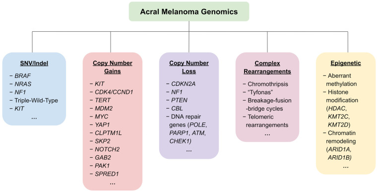
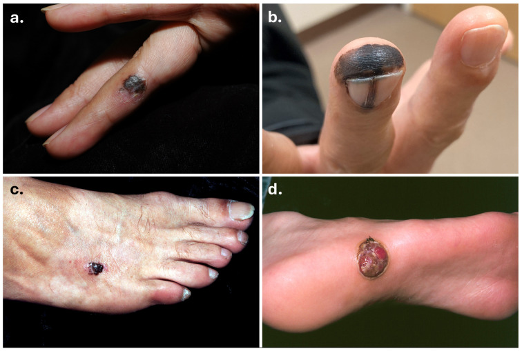
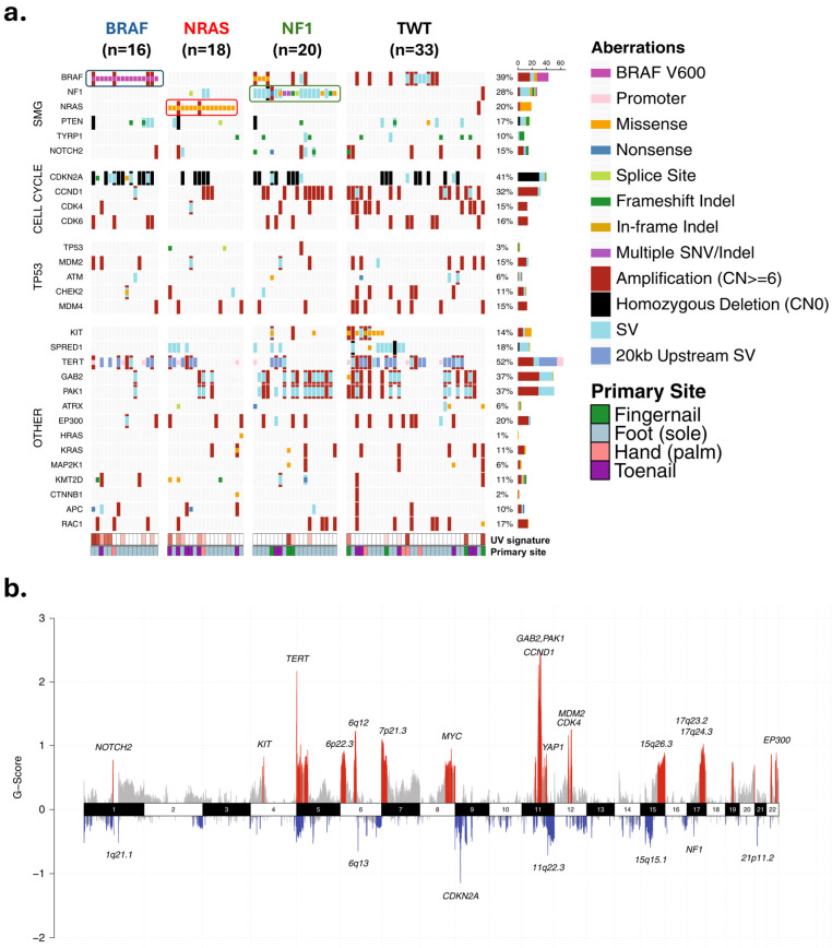
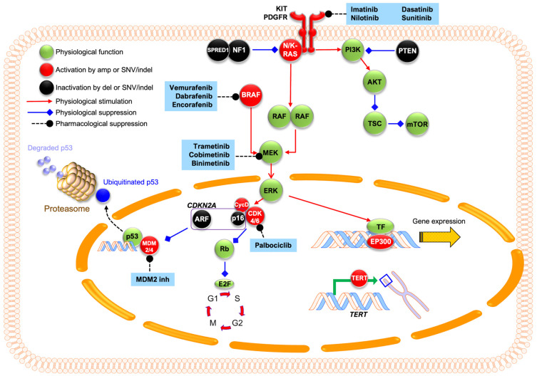
Acral melanoma is a distinct subtype of cutaneous malignant melanoma that uniquely occurs on ultraviolet (UV)-shielded, glabrous skin of the palms, soles, and nail beds. While acral melanoma only accounts for 2-3% of all melanomas, it represents the most common subtype among darker-skinned, non-Caucasian individuals. Unlike other cutaneous melanomas, acral melanoma does not arise from UV radiation exposure and is accordingly associated with a relatively low tumor mutational burden. Recent advances in genomic, transcriptomic, and epigenomic sequencing have revealed genetic alterations unique to acral melanoma, including novel driver genes, high copy number variations, and complex chromosomal rearrangements. This review synthesizes the current knowledge on the clinical features, epidemiology, and treatment approaches for acral melanoma, with a focus on the genetic pathogenesis that gives rise to its unique tumor landscape. These findings highlight a need to deepen our genetic and molecular understanding to better target this challenging subtype of melanoma.
Keywords: acral melanoma; genetics; genomics; therapy.
Conflict of interest statement
The authors declare no conflicts of interest.
Figures
Similar articles
-
Acral and nail melanoma.Clin Dermatol. 2025 Jan-Feb;43(1):3-9. doi: 10.1016/j.clindermatol.2025.01.008. Epub 2025 Feb 1. Clin Dermatol. 2025. PMID: 39900307 Review.
-
Acral melanoma: new insights into the immune and genomic landscape.Neoplasia. 2023 Dec;46:100947. doi: 10.1016/j.neo.2023.100947. Epub 2023 Oct 31. Neoplasia. 2023. PMID: 37913653 Free PMC article. Review.
-
Genetic Alterations in Primary Acral Melanoma and Acral Melanocytic Nevus in Korea: Common Mutated Genes Show Distinct Cytomorphological Features.J Invest Dermatol. 2018 Apr;138(4):933-945. doi: 10.1016/j.jid.2017.11.017. Epub 2017 Nov 27. J Invest Dermatol. 2018. PMID: 29191620
-
Clinical features, molecular pathology, and immune microenvironmental characteristics of acral melanoma.J Transl Med. 2022 Aug 16;20(1):367. doi: 10.1186/s12967-022-03532-2. J Transl Med. 2022. PMID: 35974375 Free PMC article. Review.
-
More than just acral melanoma: the controversies of defining the disease.J Pathol Clin Res. 2021 Nov;7(6):531-541. doi: 10.1002/cjp2.233. Epub 2021 Jul 2. J Pathol Clin Res. 2021. PMID: 34213090 Free PMC article. Review.
References
-
- Basurto-Lozada P., Molina-Aguilar C., Castaneda-Garcia C., Vázquez-Cruz M.E., Garcia-Salinas O.I., Álvarez-Cano A., Martínez-Said H., Roldán-Marín R., Adams D.J., Possik P.A., et al. Acral Lentiginous Melanoma: Basic Facts, Biological Characteristics and Research Perspectives of an Understudied Disease. Pigment. Cell Melanoma Res. 2021;34:59–71. doi: 10.1111/pcmr.12885. - DOI - PMC - PubMed
Publication types
MeSH terms
LinkOut - more resources
Full Text Sources
Medical
