. 2025 Jan 15;15(1):132.
doi: 10.3390/biom15010132.

From Waste to Innovation: A Circular Economy Approach for Tissue Engineering by Transforming Human Bone Waste into Novel Collagen Membranes

Affiliations

From Waste to Innovation: A Circular Economy Approach for Tissue Engineering by Transforming Human Bone Waste into Novel Collagen Membranes

Lorena Gallego et al. Biomolecules. .

Abstract

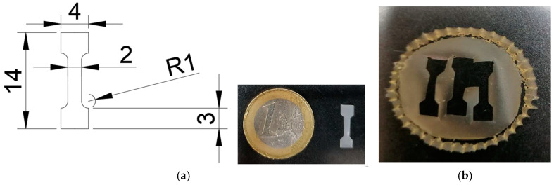
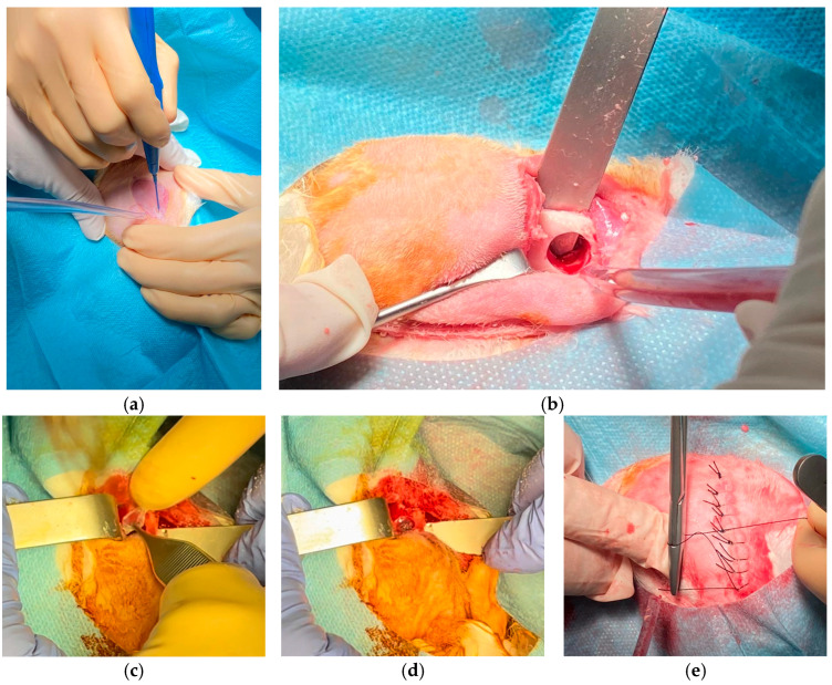
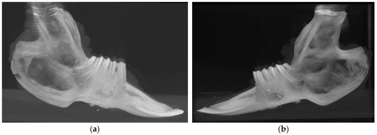
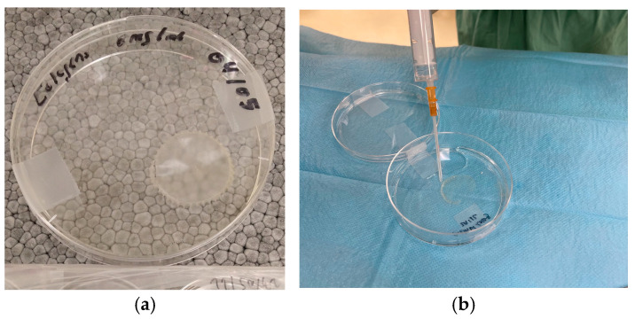
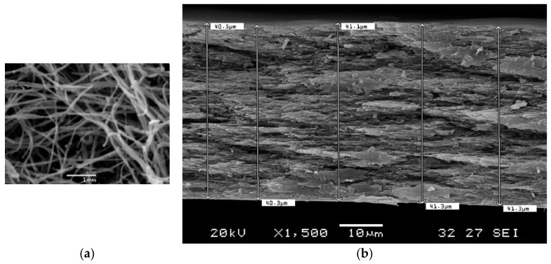
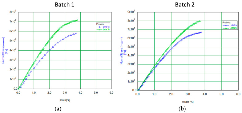
The aim of the circular economy is to treat waste as a valuable raw material, reintegrating it into the industrial economy and extending the lifecycle of subsequent products. Efforts to reduce the production of hard-to-recycle waste are becoming increasingly important to manufacturers, not only of consumer goods but also of specialized items that are difficult to manufacture, such as medical supplies, which have now become a priority for the European Union. The purpose of the study is to manufacture a novel human-purified type I collagen membrane from bone remnants typically discarded during the processing of cortico-cancellous bones in tissue banks and to evaluate its mechanical properties and effectiveness in regenerating bone-critical mandibular defects in rabbits. To prepare the novel membrane, cortico-cancellous bone chip samples from a local tissue bank were processed to isolate collagen by demineralization under agitation in HCl, cast into a silicone mold, and air-dried at room temperature and UV irradiation. The average thickness of the four batches analyzed by SEM was 37.3 μm. The average value of Young's modulus and tensile strength obtained from the specimens was 2.56 GPa and 65.43 Mpa, respectively. The membrane's efficacy was tested by creating a critical bicortical and bilateral osteoperiosteal defect in rabbit mandibles. The right-side defects were covered with the collagen membrane, while the left-side defects were left untreated as a control. Nine weeks post-surgery, clinical, radiological, and histological analyses demonstrated new bone formation in the treated areas, whereas the control sites showed no bone regeneration. This innovative approach not only contributes to sustainability in healthcare by optimizing biological waste but also exemplifies efficient resource use in line with the circular economy, offering a cost-effective, biocompatible option that could benefit national health systems.

Keywords: biodegradable polymers; bone regeneration; circular economy; collagen membrane.


Conflict of interest statement

The authors declare no conflicts of interest.

Figures

Figure 1
Figure 1
Design and dimensions of a specimen intended for mechanical tensile testing. (a) Technical drawing showing the geometry of the specimen, with measurements marked in millimeters. It has a central narrow section with a width of 2 mm and a radius of curvature (R1) at each transition point. The upper and lower sections are wider, measuring 4 mm, with a total height of 14 mm. This specimen’s design ensures that stress will concentrate in the narrow central section during mechanical testing. It also shows a physical specimen placed next to a one-euro coin, providing a sense of scale. (b) Macroscopic appearance of the membrane fabricated with the specimens used for mechanical testing.
Figure 2
Figure 2
Rabbit surgery. (a) Mandibular approach with an incision from the chin to the midpoint between mandibular angles for bilateral mandible exposure. (b) Mandibular defect created using a 10 mm trephine, perforating both cortical layers without damaging soft tissue. (c) Placement of collagen membranes on vestibular and lingual cortices to isolate the defect. (d) Membranes applied without fixation, resembling ocular lenses in flexibility. (e) Closure of muscle and skin layers with resorbable and silk sutures, respectively.
Figure 3
Figure 3
Computed tomography image of complete jaws. (a) Bone regeneration observed on the right side of the animal, where the defect was covered with a membrane. (b) Persistent defect on the left side of the animal (control side), where no membrane was applied.
Figure 4
Figure 4
Macroscopic appearance of the membrane fabricated. (a) Newly fabricated membrane showing a whitish-transparent color. (b) Hydration of the membrane with physiological saline; as shown, it acquires a transparent appearance similar to that of a contact lens.
Figure 5
Figure 5
Scanning electron microscope image of the fabricated membranes. (a) showing the fibrous structure collagen membrane. (b) Example of thickness measurement of the samples.
Figure 6
Figure 6
Stress-strain curves for the specimens from batches (a) 1 and (b) 2.
Figure 7
Figure 7
Stress-strain curves for the specimens from batches (a) 3 and (b) 4.
Figure 8
Figure 8
Computed tomography image of complete jaws. Image resolution: 31.87 µm/pixel. No magnification was applied. (a) Axial CT view highlighting anatomical regions with a defect observed on the left side and evidence of regeneration on the right. (b) Sagittal CT view with a persistent defect on the left side of the animal (control side), where no membrane was applied. (c) Sagittal CT view with bone regeneration on the right side of the animal, where the defect was covered with a membrane. (df) Same images as from (ac) but with a color palette representing different bone densities, where blue indicates the lowest density (hypodensity).
Figure 9
Figure 9
Multiple cross-sectional micro-CT views of a bone regeneration site. (a) longitudinal view, capturing both cortical and trabecular bone regions. (b) axial and (c) sagittal perspectives of the regenerated bone matrix, providing detailed insight into the bone formation and structural integration within the zone.
Figure 10
Figure 10
(a) Mean BMD variable. (b) BV/TV ratio.
Figure 11
Figure 11
Box-and-whisker plot graphic of trabecular bone comparison by type of defect. (a) Trabecular Thickness. (b) Trabecular Number. (c) Trabecular Separation.
Figure 12
Figure 12
Macroscopic appearance of a bilateral view of mandible defect healing. (a) Right side of the mandible, where the defect was treated with a membrane, bone regeneration appears to be complete, demonstrating effective structural healing. (b) The left side, which was left untreated, still displays the unhealed defect, indicating the critical nature of the defect.
Figure 13
Figure 13
Histological analysis of covered defects observed by conventional light microscope stained with Masson’s trichrome (ae) and Vimentin (f).

Similar articles

References

    1. Manyalich M., Navarro A., Koller J., Loty B., De Guerra A., Cornu O., Vabels G., Fornasari P.M., Costa A.N., Siska I., et al. European Quality System for Tissue Banking. Transplant. Proc. 2009;41:2035–2043. doi: 10.1016/j.transproceed.2009.06.157. - DOI - PubMed
    1. Organización Nacional de Trasplantes; Asociación Española de Bancos de Tejidos Plan Estratégico Nacional de Tejidos 2022-2026. 2022. [(accessed on 15 September 2024)]. Available online: https://www.ont.es/wp-content/uploads/2023/06/PLAN-ESTRATEGICO-NACIONAL-....
    1. Howkins J. ReThinking the Creative Economy. 2022. [(accessed on 20 November 2024)]. Available online: https://www.academia.edu/82397309/ReThinking_the_Creative_Economy.
    1. Dziuba R., Kucharska M., Madej-Kiełbik L., Sulak K., Wiśniewska-Wrona M. Biopolymers and Biomaterials for Special Applications within the Context of the Circular Economy. Materials. 2021;14:7704. doi: 10.3390/ma14247704. - DOI - PMC - PubMed
    1. Aguilar A., Twardowski T., Wohlgemuth R. Bioeconomy for Sustainable Development. Biotechnol. J. 2019;14:1800638. doi: 10.1002/biot.201800638. - DOI - PubMed

LinkOut - more resources