. 2025 Jan 20;15(1):150.
doi: 10.3390/biom15010150.

Cell Type Specific Suppression of Hyper-Recombination by Human RAD18 Is Linked to Proliferating Cell Nuclear Antigen K164 Ubiquitination

Affiliations

Cell Type Specific Suppression of Hyper-Recombination by Human RAD18 Is Linked to Proliferating Cell Nuclear Antigen K164 Ubiquitination

Colette B Rogers et al. Biomolecules. .

Abstract

RAD18 is a conserved eukaryotic E3 ubiquitin ligase that promotes genome stability through multiple pathways. One of these is gap-filling DNA synthesis at active replication forks and in post-replicative DNA. RAD18 also regulates homologous recombination (HR) repair of DNA breaks; however, the current literature describing the contribution of RAD18 to HR in mammalian systems has not reached a consensus. To investigate this, we examined three independent RAD18-null human cell lines. Our analyses found that loss of RAD18 in HCT116, but neither hTERT RPE-1 nor DLD1 cell lines, resulted in elevated sister chromatid exchange, gene conversion, and gene targeting, i.e., HCT116 mutants were hyper-recombinogenic (hyper-rec). Interestingly, these phenotypes were linked to RAD18's role in PCNA K164 ubiquitination, as HCT116 PCNAK164R/+ mutants were also hyper-rec, consistent with previous studies in rad18-/- and pcnaK164R avian DT40 cells. Importantly, the knockdown of UBC9 to prevent PCNA K164 SUMOylation did not affect hyper-recombination, strengthening the link between increased recombination and RAD18-catalyzed PCNA K164 ubiquitination, but not K164 SUMOylation. We propose that the hierarchy of post-replicative repair and HR, intrinsic to each cell type, dictates whether RAD18 is required for suppression of hyper-recombination and that this function is linked to PCNA K164 ubiquitination.

Keywords: PCNA K164; RAD18; gap-filling; hyper-recombination; ubiquitination.


Conflict of interest statement

A.K.B. is a member of the editorial board of Biomolecules. D.A.L. is the co-founder and co-owner of several biotechnology companies including NeoClone Biotechnologies, Inc., Discovery Genomics, Inc. (recently acquired by Immusoft, Inc.), B-MoGen Biotechnologies, Inc. (recently acquired by Bio-Techne Corporation), and Luminary Therapeutics, Inc. D.A.L. holds equity in, serves as a Senior Scientific Advisor for and Board of Director member for Recombinetics, a genome editing company. D.A.L. consults for Genentech, Inc., which is funding some of his research. The business of all these companies is unrelated to the contents of this manuscript.

Figures

Figure 1
Figure 1
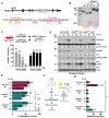
RAD18-mediated suppression of recombination is cell line specific. (AC) Western blot analyses confirm the loss of RAD18 in two independent RPE-1 (A), DLD1 (B), and HCT116 (C) RAD18−/− clones. The top band detected by the anti-RAD18 antibody is mono-ubiquitinated RAD18 and the bottom band is unmodified RAD18 [45]. The asterisk indicates a nonspecific band recognized by the anti-RAD18 antibody. Quantification is based on densitometry using FIJI normalized to the loading control and WT lane, minus signal from the nonspecific RAD18 band. (D) Schematic of the SCE assay. (E) Example metaphase spread from an HCT116 RAD18−/− cell used for SCE analyses. One SCE is scored each time there is a switch from green-labeled DNA to blue-labeled DNA (or vice versa) on a sister chromatid. The scale bar is 10 µm. (FH) The average number of SCEs per chromosome in RPE-1 (F), DLD1 (G), or HCT116 (H) cell lines. Each dot represents one metaphase spread across two biological replicates. Black lines indicate mean values and significance was calculated using a Mann–Whitney test with **** > 0.0001 and ns = not significant. (I) Western blot analysis confirming complementation of HCT116 RAD18−/− clone 1A3 with wild-type FLAG-tagged RAD18 in two independent clones (2.3 and 2.4). The asterisk indicates a nonspecific band recognized by the anti-RAD18 antibody. Quantification is based on densitometry using FIJI normalized to the loading control and WT lane (RAD18) minus signal from the nonspecific RAD18 band or normalized to the loading control and 2.3 lane (FLAG). (J) Average number of SCEs per chromosome in HCT116 cell lines. Each dot represents one metaphase spread within a single biological replicate. Black lines indicate mean values and significance was calculated using a Mann–Whitney test with **** > 0.0001. Original western blot images can be found in Figure S2.
Figure 2
Figure 2
HCT116 RAD18/ cells exhibit hyper-recombination phenotypes. (A) Schematic of the DR-GFP gene conversion reporter assay. (BD) Results of the DR-GFP gene conversion assay. The average percentage of GFP+ cells normalized to wild-type for RPE-1 (B), DLD1 (C) or HCT116 (D) cell lines. For each cell line n = at least 9 across at least three biological replicates. Each column is normalized to the baseline, defined as the mean of all WT values. The error is indicated as standard deviation and significance was calculated using Dunnett’s one-way ANOVA tests, with ** > 0.01 and ns = not significant. (E) Schematic of PIGA gene targeting assay. (F,G) Results of the PIGA gene targeting assay. The average percentage of FLAER-positive (FLAER+) cells in HCT116 (F) or RPE-1 (G) cell lines. For each cell line n = 9 wells across three biological replicates. The error is indicated as standard deviation and significance was calculated using Dunnett’s one-way ANOVA tests, with * > 0.05 and ns = not significant.
Figure 3
Figure 3
TLS activation is reduced in RAD18/ cells without significantly decreased viability following UV irradiation. (A) Western blot analysis of POL η, RAD18, PCNA, and PCNA ubiquitinated at K164 levels in nuclear fractions from HCT116 wild-type and RAD18/ cell lines with or without preceding UV irradiation (10 or 20 J). The asterisk indicates a nonspecific band recognized by the anti-RAD18 antibody. Quantification is based on densitometry using FIJI normalized to the loading control and WT lane. (B) Clonogenic survival of HCT116 wild-type and two RAD18/− cell lines following UV irradiation at the indicated dosages (5, 10, or 20 J). Error is indicated as SEM and significance was calculated using an unpaired, two-tailed Student’s t-test across four biological replicates. n.s. = not significant. Original western blot images can be found in Figure S3.
Figure 4
Figure 4
Generation and characterization of HCT116 PCNAK164R/+ mutant cell lines. (A) Schematic of the PCNA exon 5 targeting strategies used to generate HCT116 PCNAK164R/+ mutant cell lines. (B) Representative results from the diagnostic PCR followed by EcoRI or XcmI restriction enzyme digestion to identify successful targeting of PCNA exon 5. Expected products for the 5′ targeting strategy include the following: not targeted (WT) 180 bp, 1246 bp; biallelic targeting (PCNAK164R/+ 1A2, 1D10) 180 bp, 992 bp, and 254 bp. Expected products for the 3′ targeting strategy include the following: not targeted (WT) 1460 bp; biallelic targeting (PCNAK164R/+ 1B9) 1260 bp and 166 bp. (C) (Top) Schematic of an experiment to determine HCT116 PCNA haplolethality. (Bottom) The percent INDEL frequency as measured by TIDE analysis in HCT116 populations following PCNA exon 2 or MCM10 exon 3 CRISPR/Cas9 targeting, respectively, over a seven-day time course. Error is indicated as standard deviation representing two biological replicates. (D) Western blot analyses of PCNA, ubiquityl-PCNA (K164), phospho-RPA32 (S4/8), and γH2AX, with or without UV treatment (20 or 40 J), with histone H2AX as the loading control, in nuclear fractions isolated from HCT116 cell lines. Quantification is based on densitometry using FIJI normalized to the loading control and WT lane. (E) Clonogenic survival of HCT116 wild-type and two PCNAK164R/+ cell lines following UV irradiation at the indicated dosages (5, 7, or 10 J). Error is indicated as standard deviation and significance was calculated using a two-tailed students t-test with * > 0.05; ** > 0.01 across at least three biological replicates. (F) Schematic of the HPRT survival assay. (G) Average mutation frequency at the HPRT locus in untreated or UV-treated (6 J) HCT116 cell lines. Error is indicated as SEM and significance was calculated using Sidak’s multiple comparison test across three biological replicates. n.s. = not significant. No comparisons were statistically significant. Original western blot images can be found in Figure S5.
Figure 5
Figure 5
Reduced PCNA K164 ubiquitination leads to increased global and telomeric recombination. (A) Western blot analysis of PCNA levels in nuclear fractions with or without UV treatment (20 or 40 J), with histone H2AX as the loading control, in HCT116 wild-type, PCNAK164R/+, and PCNA+/+ reverted cell lines. Quantification is based on densitometry using FIJI normalized to the loading control and WT lane. (B) Quantification of the average number of SCEs per chromosome in HCT116 wild-type, PCNAK164R/+, and PCNA+/+ reverted ST cell lines. Black lines indicate mean values and statistical significance was calculated using Kruskal–Wallis with Dunn’s multiple comparison test with *** < 0.001; across two biological replicates. (C) Representative metaphase spread for spontaneous telomeric SCEs (t-SCEs). Examples of a t-SCE event and chromosomes without t-SCE are highlighted. The scale bar is 20 µm. (D) Quantification of the percent t-SCEs per chromosome ends in HCT116 wild-type, PCNAK164R/+, and PCNA+/+ reverted ST cell lines. Black lines indicate mean values and statistical significance was calculated using Kruskal–Wallis with Dunn’s multiple comparison test *** < 0.001; across two biological replicates. (E) Western blot analysis of UBC9 and PCNA levels in HCT116 wild-type, PCNAK164R/+, and PCNA+/+ reverted ST cell lines following a 72 h treatment with siControl or siUBC9, with tubulin as a loading control. Quantification is based on densitometry using FIJI normalized to the loading control and WT lane. (F) Quantification of the percent t-SCEs per chromosome ends in HCT116 wild-type, PCNAK164R/+, and PCNA+/+ reverted ST cell lines following a 72 h treatment with siControl or siUBC9. Black lines indicate mean values and statistical significance was calculated using Kruskal–Wallis with Dunn’s multiple comparison test ** < 0.01, **** < 0.0001, n.s. = not significant; across two biological replicates. Original western blot images can be found in Figure S6.
Figure 6
Figure 6
Suppression of hyper-recombination by RAD18 is linked to PCNA K164 ubiquitination. In wild-type HCT116 and DT40 cells, the balance between gap-filling DNA synthesis and HR is regulated by the activity of RAD18 and the level of PCNA K164 ubiquitination. In these cell lines, loss of RAD18 results in a significant increase in HR. We speculate that this occurs to compensate for the deficiency in gap-filling DNA synthesis and improve viability.

Update of

References

    1. Edmunds C.E., Simpson L.J., Sale J.E. PCNA ubiquitination and REV1 define temporally distinct mechanisms for controlling translesion synthesis in the avian cell line DT40. Mol. Cell. 2008;30:519–529. doi: 10.1016/j.molcel.2008.03.024. - DOI - PubMed
    1. Guilliam T.A., Yeeles J.T.P. Reconstitution of translesion synthesis reveals a mechanism of eukaryotic DNA replication restart. Nat. Struct. Mol. Biol. 2020;27:450–460. doi: 10.1038/s41594-020-0418-4. - DOI - PMC - PubMed
    1. Khatib J.B., Nicolae C.M., Moldovan G.L. Role of Translesion DNA Synthesis in the Metabolism of Replication-associated Nascent Strand Gaps. J. Mol. Biol. 2024;436:168275. doi: 10.1016/j.jmb.2023.168275. - DOI - PMC - PubMed
    1. Jasin M., Rothstein R. Repair of strand breaks by homologous recombination. Cold Spring Harb. Perspect. Biol. 2013;5:a012740. doi: 10.1101/cshperspect.a012740. - DOI - PMC - PubMed
    1. Wright W.D., Shah S.S., Heyer W.D. Homologous recombination and the repair of DNA double-strand breaks. J. Biol. Chem. 2018;293:10524–10535. doi: 10.1074/jbc.TM118.000372. - DOI - PMC - PubMed

MeSH terms

LinkOut - more resources