

Comparison of Lipid Content in Nine Dinoflagellate Species Using Flow Cytometry
- PMID: 39858812
- PMCID: PMC11768077
- DOI: 10.3390/microorganisms13010044
Comparison of Lipid Content in Nine Dinoflagellate Species Using Flow Cytometry
Abstract
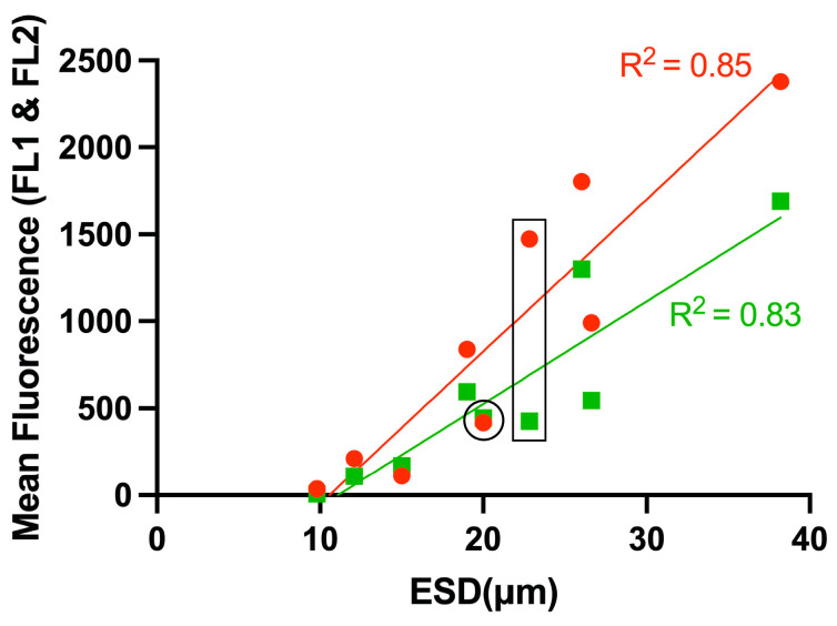
The lipid content of nine dinoflagellates was analyzed using flow cytometry to compare lipid levels. Additionally, the correlation between lipid content, cell size, and carbon content in dinoflagellates was evaluated using BODIPY 505/515 staining. The flow cytometry side scatter (SSC) effectively represented relative cell size, showing a linear relationship with the equivalent spherical diameter (ESD). Larger cells exhibited higher relative lipid content; however, lipid accumulation was influenced by nutritional modes and habitats, with mixorophic and benthic species displaying higher lipid content than heterotrophic species. A comparison of fluorescent dyes revealed that Nile Red overestimated lipid content, suggesting overlap with chlorophyll autofluorescence. Flow cytometry analysis with BODIPY 505/515 demonstrated a linear correlation with the sulfo-phospho-vanillin (SPV) method, enabling determination of actual lipid content using FL1 fluorescence and the slope value. As the carbon content increased, the lipid content initially increased rapidly but plateaued at higher carbon levels, indicating saturation. These findings suggest that relative fluorescence via flow cytometry provides an effective means to estimate the lipid content and carbon content as a function of cell size.
Keywords: carbon content; cell size; dinoflagellate; flow cytometry; lipid content.
Conflict of interest statement
The authors declare no conflicts of interest.
Figures


References
-
- Piretti M.V., Pagliuca G., Boni L., Pistocchi R., Diamante M., Gazzotti T. Investigation of 4-methyl sterols from cultured dinoflagellate algal strains. J. Phycol. 1997;33:61–67. doi: 10.1111/j.0022-3646.1997.00061.x. - DOI
-
- Mansour M.P., Volkman J.K., Jackson A.E., Blackburn S.I. The fatty acid and sterol composition of five marine dinoflagellates. J. Phycol. 1999;35:710–720. doi: 10.1046/j.1529-8817.1999.3540710.x. - DOI
-
- Muscatine L. The role of symbiotic algae in carbon and energy flux in reef corals. In: Dubinski Z., editor. Coral Reefs: Ecosystems of the World. Elsevier; New York, NY, USA: 1990. pp. 75–87.
-
- Jeong H.J., Yoo Y.D., Kim J.S., Seong K.A., Kang N.S., Kim T.H. Growth, feeding and ecological roles of mixotrophic and heterotrophic dinoflagellates in marine planktonic food webs. Ocean Sci. J. 2010;45:65–91. doi: 10.1007/s12601-010-0007-2. - DOI
-
- de la Jara A., Mendoza H., Martel A., Molina C., Nordströn L., de la Rosa V., Díaz R. Flow cytometric determination of lipid content in a marine dinoflagellate, Crypthecodinium cohnii. J. Appl. Phycol. 2003;15:433–438. doi: 10.1023/A:1026007902078. - DOI
Grants and funding
LinkOut - more resources
Full Text Sources
Research Materials
Miscellaneous
