. 2025 Jan 10;61(1):100.
doi: 10.3390/medicina61010100.

Technical Report on the New Ultrasound Lateral Mid-Shaft Approach to the Sciatic Nerve: A Never-Ending Story

Affiliations

Technical Report on the New Ultrasound Lateral Mid-Shaft Approach to the Sciatic Nerve: A Never-Ending Story

Giuseppe Sepolvere et al. Medicina (Kaunas). .

Abstract

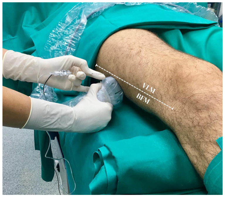
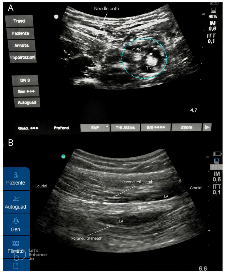
The anatomy of the sciatic nerve allows it to be blocked at different levels using various anesthetic approaches. However, for several reasons, performing these approaches may be challenging or disadvantageous in specific categories of patients, particularly in obese patients. The objective of this brief technical report is to describe a new technical approach to sciatic nerve block, designed to simplify the procedure for certain categories of patients and less experienced practitioners. Since 2010, more than 5000 procedures have been performed by both experienced anesthesiologists and novice trainees in several hospitals. The ultrasound lateral mid-shaft technique appears to be a safe and effective method for performing a sciatic nerve block, even in obese patients with significant subcutaneous fat and unclear ultrasound images. This approach is particularly beneficial given the various anatomical variations that can occur. By targeting the mid-thigh area, the ultrasound beam accesses anatomical structures that are more superficial, improving the technique's efficacy. Various hospital groups have been performing this technique as a routine procedure, achieving a success rate of nearly 100%. This impressive success rate exceeds that of other conventional techniques documented in the literature. Additionally, there have been significant improvements in comfort and ease for anesthetists. This method allows the anesthetic to spread around the paraneural sheath, covering the posterior femoral cutaneous nerve. Finally, it is performed in the supine position without the need to mobilize the lower limbs, ensuring patient comfort, especially in cases of fractures or lower limb injuries. Further studies are needed to confirm these results.

Keywords: loco-regional anesthesia; lower limb; nerve block; sciatic nerve.


Conflict of interest statement

The authors declare no conflicts of interest.

Figures

Figure 4
Figure 4
Different approaches to the sciatic nerve. (A) Popliteal approach: when performing the block, the prone position is uncomfortable in the case of lower limb fracture. (B) Lateral approach: the original lateral approach requires the patient to adopt a supine patient with their leg bent, a lateral to medial needle direction, and the probe placed below the thigh with a posterior to anterior ultrasound beam, similar to the popliteal approach performed with the patient in a supine position. The challenges consist of the uncomfortable position adopted by both the patient, unable to flex their leg, and the anesthetist. (C) Anterior approach: the patient is placed in a comfortable supine position, but the nervous target is overly deep, making it difficult for the local anesthetic to surround the nervous target by the local anesthetic and making the procedure painful. (D) Subgluteal approach: the patient is placed in a lateral position with the lower limb upward. Similar to the popliteal approach, the procedure can be uncomfortable in the case of fracture.
Figure 1
Figure 1
A convex ultrasound probe is positioned at the mid-tight in the muscular groove formed by the vastus lateralis and femoral biceps muscles (white dotted line), with the beam oriented upward. VLM: vastus lateralis muscle; BFM: biceps femoris muscle.
Figure 2
Figure 2
(A) Sciatic nerve divided into its components within the same paraneural sheath (light blue dotted circle) after the injection of local anesthetic. CPN: common peroneal nerve; TN: tibial nerve (B) Ultrasound long-axis view after lateral mid-shaft sciatic nerve block: the local anesthetic correctly injected below the paraneural sheath may spread for several centimeters in a caudal to cranial direction up to the subgluteal region covering the posterior femoro-cutaneous branch. LA: local anesthetic; SN: sciatic nerve.
Figure 3
Figure 3
New ultrasound lateral mid-shaft approach to the SN: step by step flowchart.
Figure 5
Figure 5
The presence of a vascular structure could be visualized within the body of the sciatic nerve. In the picture, the doppler shows the presence of the artery on the short axis inside the body of the sciatic nerve. SN: sciatic nerve; LA: local anesthetic.

References

    1. Vadivelu N., Kai A.M., Maslin B., Kodumudi V., Antony S., Blume P. Role of regional anesthesia in foot and ankle surgery. Foot Ankle Spec. 2015;8:212–219. doi: 10.1177/1938640015569769. - DOI - PubMed
    1. Shevlin S., Johnston D., Turbitt L. The sciatic nerve block. BJA Educ. 2020;20:312–320. doi: 10.1016/j.bjae.2020.04.004. - DOI - PMC - PubMed
    1. Shoja M.M. Pelvic girdle, gluteal region and thigh. In: Standring S., editor. Gray’s Anatomy: The Anatomical Basis of Clinical Practice. 41st ed. Elsevier; New York, NY, USA: 2016.
    1. Karmakar M.K., Shariat A.N., Pangthipampai P., Chen J. High-definition ultrasound imaging defines the paraneural sheath and the fascial compartments surrounding the sciatic nerve at the popliteal fossa. Reg. Anesth. Pain Med. 2013;38:447–451. doi: 10.1097/AAP.0b013e31829ffcb4. - DOI - PubMed
    1. Perlas A., Wong P., Abdallah F., Hazrati L.N., Tse C., Chan V. Ultrasound-guided popliteal block through a common paraneural sheath versus conventional injection: A prospective, randomized, double-blind study. Reg. Anesth. Pain Med. 2013;38:218–225. doi: 10.1097/AAP.0b013e31828db12f. - DOI - PubMed

MeSH terms

LinkOut - more resources