

Identification of Biomarkers of Arrhythmogenic Cardiomyopathy (ACM) by Plasma Proteomics
- PMID: 39859087
- PMCID: PMC11766713
- DOI: 10.3390/medicina61010105
Identification of Biomarkers of Arrhythmogenic Cardiomyopathy (ACM) by Plasma Proteomics
Abstract
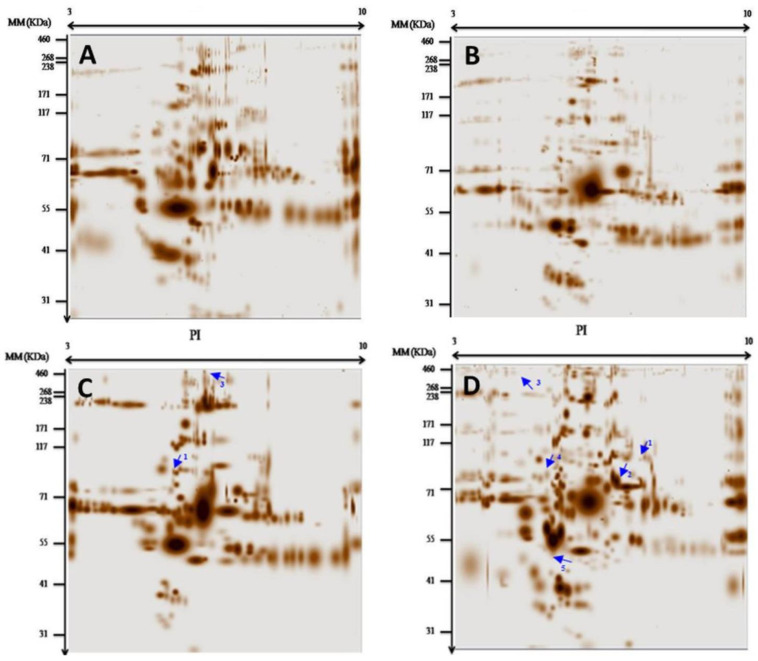
Background and Objectives: The pathophysiology of arrhythmogenic cardiomyopathy (ACM), previously known as arrhythmogenic right ventricular cardiomyopathy (ARVC), and its specific biological features remain poorly understood. High-throughput plasma proteomic profiling, a powerful tool for gaining insights into disease pathophysiology at the systems biology level, has not been used to study ACM. This study aimed at characterizing plasmatic protein changes in patients with ACM, which were compared with those of healthy controls, and at exploring the potential role of the identified proteins as biomarkers for diagnosis and monitoring. Materials and Methods: Blood samples were collected from six ACM patients, four patients with other cardiomyopathies, and two healthy controls. Plasma was processed to remove high-abundance proteins and analyzed by two-dimensional gel electrophoresis. Differential protein expressions were assessed using PDQuest software, Bio-Rad US version 8.0.1. Results: The analysis revealed several proteins with altered expressions between ACM patients and controls, including plakophilin-2, junctional plakoglobin, desmoplakin, desmin, transmembrane protein 43, and lamin A/C. Conclusions: The plasma proteomic profiling of ACM suggests that ACM is a distinct disease entity characterized by a unique dysregulation of desmosomal proteins. The identification of plasma biomarkers associated with ACM underscores their potential to improve diagnostic accuracy and facilitate early intervention strategies. Further exploration of mutations in desmosomal proteins and their phosphorylation states may provide deeper insights into the pathophysiology of ACM.
Keywords: arrhythmogenic cardiomyopathy; biomarkers; desmosomal proteins; phosphorylation; proteomics.
Conflict of interest statement
The authors declare no conflict of interest.
Figures

References
-
- Tabib A., Loire R., Chalabreysse L., Meyronnet D., Miras A., Malicier D., Thivolet F., Chevalier P., Bouvagnet P. Circumstances of death and gross and microscopic observations in a series of 200 cases of sudden death associated with arrhythmogenic right ventricular cardiomyopathy and/or dysplasia. Circulation. 2003;108:3000–3005. doi: 10.1161/01.CIR.0000108396.65446.21. - DOI - PubMed
-
- Basso C., Czarnowska E., Barbera M.D., Bauce B., Beffagna G., Wlodarska E.K., Pilichou K., Ramondo A., Lorenzon A., Wozniek O., et al. Ultrastructural evidence of intercalated disc remodelling in arrhythmogenic right ventricular cardiomyopathy: An electron microscopy investigation on endomyocardial biopsies. Eur. Heart J. 2006;27:1847–1854. doi: 10.1093/eurheartj/ehl095. - DOI - PubMed
MeSH terms
Substances
LinkOut - more resources
Full Text Sources
