. 2025 Jan 8;26(2):458.
doi: 10.3390/ijms26020458.

Potentiation of Gelonin Cytotoxicity by Pulsed Electric Fields

Affiliations

Potentiation of Gelonin Cytotoxicity by Pulsed Electric Fields

Olga N Pakhomova et al. Int J Mol Sci. .

Abstract

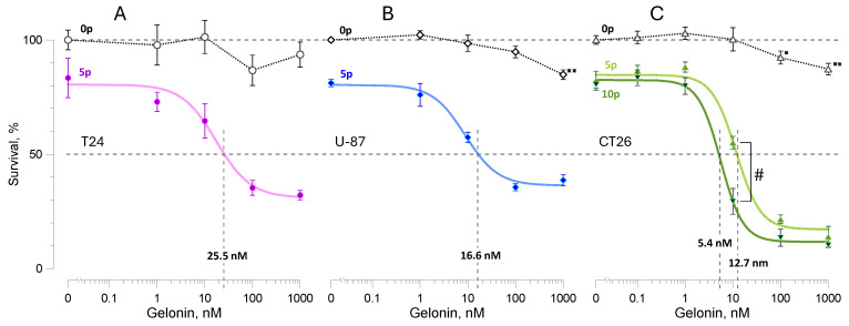
Gelonin is a ribosome-inactivating protein with extreme intracellular toxicity but poor permeation into cells. Targeted disruption of cell membranes to facilitate gelonin entry is explored for cancer and tissue ablation. We demonstrate a hundreds- to thousands-fold enhancement of gelonin cytotoxicity by pulsed electric fields in the T24, U-87, and CT26 cell lines. The effective gelonin concentration to kill 50% of cells (EC50) after electroporation ranged from <1 nM to about 100 nM. For intact cells, the EC50 was unattainable even at the highest gelonin concentration of 1000 nM, which reduced cell survival by only 5-15%. For isoeffective electroporation treatments using 300 ns, 9 µs, and 100 µs pulses, longer pulses were more efficient at lowering gelonin EC50. Increasing the electric field strength of 8, 100 µs pulses from 0.65 to 1.25 kV/cm reduced gelonin EC50 from 128 nM to 0.72 nM. Conversely, the presence of 100 nM gelonin enabled a more than 20-fold reduction in the number of pulses required for equivalent cell killing. Pulsed electric field-mediated delivery of gelonin shows promise for hyperplasia ablation at concentrations sufficiently low to minimize or avoid systemic toxicity.

Keywords: cancer ablation; electrochemotherapy; electropermeabilization; electroporation; irreversible electroporation; pulsed field ablation.


Conflict of interest statement

The authors declare no conflicts of interest except the patent pending for A.G.P and O.P. The funders had no role in the design of the study; in the collection, analyses, or interpretation of data; in the writing of the manuscript; or in the decision to publish the results.

Figures

Figure 1
Figure 1
Potentiation of gelonin cytotoxicity by pulsed electric fields in T24 (A), U-87 (B), and CT26 (C) cell lines. Viability was measured 24 h after electroporation with 9-µs, 2 kV/cm pulses at 10 Hz. Samples were treated with 5 pulses (“5p”, AC) or 10 pulses (“10p”, C) in the presence of gelonin at 0.1–1000 nM or the vehicle (0 nM). Control samples (open symbols) underwent the same treatment, but no electric pulses were delivered (sham exposure, “0p”). Viability in the 0p group at 0 nM gelonin was used as the 100% reference. Solid color lines are best fits for the 5p and 10p groups using the Hill equation. The effective gelonin concentrations that reduced viability to 50% (EC50) were measured from Hill fits and are shown by vertical dashed lines and legends above the x-axis. Mean ± s.e., n = 4–7. * p < 0.05, ** p < 0.01 for the difference of sham-exposed controls from 100% (one sample, 2-tailed t-test). # p < 0.01 for the difference between the 5p and 10p groups (unpaired 2-tailed t-test). Differences in viability of the electroporated and sham-exposed cells are significant at p < 0.05 or better for all gelonin concentrations (not labeled in the graphs).
Figure 2
Figure 2
The effect of gelonin and electroporation on the viability change from 24 to 48 h in CT26 cells. Samples were treated with 5 (A) or 10 (B) electric pulses (9 µs, 2 kV/cm) or were sham-exposed in the presence of different gelonin concentrations (0.001–1000 nM) or vehicle (0 nM). Viability measured at 24 h in each individual sample was taken as 100%. Mean ± s.e., n = 4; * p < 0.05, ** p < 0.01 for the difference from the matching sham-exposed control group (unpaired 2-tailed t-test). See Figure 1 and the text for more detail.
Figure 3
Figure 3
Effect of the electric pulse duration on the potentiation of gelonin cytotoxicity in CT26 cells. Doses of 300-ns, 9-µs, and 100-µs pulses (legends) were adjusted to cause a similar reduction in viability (to 80–90%) in the absence of gelonin. Gelonin EC50 values (legends above x-axis) were smaller in cells electroporated by longer electric pulses. Mean ± s.e., n = 4 for electric pulse-treated groups. Data for sham-exposed controls (open symbols, 0 kV/cm) were pooled together for all pulse durations, n = 12. # p < 0.05 and ## p < 0.01 for the difference between the samples treated with 100 µs pulses from those treated with either 9 µs or 300 ns pulses (unpaired 2-tailed t-test with Dunnett’s correction). * p < 0.05 for the difference of sham-exposed controls from 100% (one sample, 2-tailed t-test). See Figure 1 and the text for other details.
Figure 4
Figure 4
The effect of the electric field strength on the potentiation of gelonin cytotoxicity. (A) Viability measured 24 h after exposure to 8, 100-µs pulses at the indicated electric field strengths (kV/cm) in the presence of gelonin at 0.1–1000 nM or the vehicle (0 nM). Control samples (open symbols) were sham exposed (0 kV/cm). EC50 values for gelonin (nM) are labeled at the intersections of the Hill fits (solid lines) with a grid line at 50%. (B) The same data re-scaled between the viability of electroporated cells without gelonin (100%) and the observed viability minimum (0%). Mean ± s.e., n = 4 for pulse-treated groups and n = 16 for the pooled sham-exposed group. # p < 0.01 for the difference of the 1.25 kV/cm group from the 0.9 and 0.65 kV/cm groups; $ p < 0.05 for the difference of the 1.25 kV/cm group from all other groups (unpaired 2-tailed t-test with Dunnett’s correction). See Figure 1 and the text for other details.
Figure 5
Figure 5
Gelonin potentiates cell inactivation by electroporation. Viability of CT26 cells was measured 24 h after exposure to different numbers of 9-µs, 2 kV/cm pulses, in the presence or absence of 100 nM gelonin (filled and open symbols, respectively). Viability in sham-exposed samples (0 pulses) without the drug was taken as 100%. Solid and dotted lines are the respective best fits of data using the power function; the initial plateau in the no-drug group was excluded from the fit. Mean ± s.e., n = 3–4. The difference in viability with and without gelonin is significant at least at p < 0.02 (unpaired 2-tailed t-test) for all pulse numbers. Dashed lines illustrate the estimated 50-fold difference in the pulse number to achieve the same viability reduction with and without gelonin.

References

    1. Flavell D.J., Flavell S.U. Plant-Derived Type I Ribosome Inactivating Protein-Based Targeted Toxins: A Review of the Clinical Experience. Toxins. 2022;14:563. doi: 10.3390/toxins14080563. - DOI - PMC - PubMed
    1. Walsh M.J., Dodd J.E., Hautbergue G.M. Ribosome-inactivating proteins: Potent poisons and molecular tools. Virulence. 2013;4:774–784. doi: 10.4161/viru.26399. - DOI - PMC - PubMed
    1. Stirpe F. Ribosome-inactivating proteins: From toxins to useful proteins. Toxicon. 2013;67:12–16. doi: 10.1016/j.toxicon.2013.02.005. - DOI - PubMed
    1. Bolognesi A., Bortolotti M., Maiello S., Battelli M.G., Polito L. Ribosome-Inactivating Proteins from Plants: A Historical Overview. Molecules. 2016;21:1627. doi: 10.3390/molecules21121627. - DOI - PMC - PubMed
    1. Schlaak L., Weise C., Kuropka B., Weng A. Mutational Analysis of RIP Type I Dianthin-30 Suggests a Role for Arg24 in Endocytosis. Toxins. 2024;16:219. doi: 10.3390/toxins16050219. - DOI - PMC - PubMed

Substances

LinkOut - more resources