. 2025 Jan 9;26(2):490.
doi: 10.3390/ijms26020490.

Synthetic GPR84 Agonists in Colorectal Cancer: Effective in THP-1 Cells but Ineffective in BMDMs and MC38 Mouse Tumor Models

Affiliations

Synthetic GPR84 Agonists in Colorectal Cancer: Effective in THP-1 Cells but Ineffective in BMDMs and MC38 Mouse Tumor Models

Marlene Schwarzfischer et al. Int J Mol Sci. .

Abstract

Tumor-associated macrophages (TAMs) in the colorectal cancer (CRC) microenvironment promote tumor progression but can be reprogrammed into a pro-inflammatory state with anti-cancer properties. Activation of the G protein-coupled receptor 84 (GPR84) is associated with pro-inflammatory macrophage polarization, making it a potential target for CRC therapy. This study evaluates the effects of the GPR84 agonists 6-OAU and ZQ-16 on macrophage activation and anti-cancer efficacy. GPR84 expression on THP-1 macrophages and murine BMDMs was analyzed using flow cytometry. Macrophages were treated with 6-OAU or ZQ-16, and pro-inflammatory cytokine levels, reactive oxygen species (ROS) production, and phagocytosis were assessed using qPCR and functional assays. Anti-cancer effects were tested in a subcutaneous MC38 tumor model, with oral or intraperitoneal agonist administration. Pharmacokinetics and compound stability were also evaluated. In THP-1 macrophages, 6-OAU increased pro-inflammatory cytokines and ROS production, with ZQ-16 showing similar effects. However, neither agonist induced pro-inflammatory responses, ROS production, or phagocytosis in murine macrophages. In vivo, both agonists failed to inhibit tumor growth in the MC38 model despite systemic exposure. Current GPR84 agonists lack efficacy in promoting anti-cancer macrophage activity, limiting their potential as CRC therapies.

Keywords: 6-OAU; GPR84; ZQ-16; anti-cancer therapy; colorectal cancer (CRC); immunotherapy; macrophage activation; tumor-associated macrophages (TAMs).


Conflict of interest statement

The authors declare no conflicts of interest.

Figures
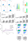
Figure 1
Figure 1
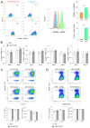
In THP-1 cells, LPS increased GPR84 expression, and 6-OAU enhanced pro-inflammatory cytokine transcription. (A) Flow cytometry of GPR84 surface and mRNA expression after LPS stimulation (100 ng/mL, 16 h). Bar charts quantify expression levels. (B) mRNA expression of GPR84 targets after LPS (0–100 ng/mL) and 6-OAU (1 µM) stimulation. (C) ROS production kinetics in response to 6-OAU (0.01–10 µM) treatment. (D) Phagocytosis of opsonized beads with LPS (100 ng/mL, 16 h) and 6-OAU (1 µM, 15–120 min) treatment. Statistical analysis: (A) Student’s t-test; (BD) one-way ANOVA and Tukey’s test, with * p ≤ 0.05, ** p ≤ 0.01, *** p ≤ 0.001, and **** p ≤ 0.0001. n = 3.
Figure 2
Figure 2
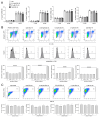
In murine BMDMs, LPS increased GPR84 expression, but 6-OAU did not enhance pro-inflammatory signaling or phagocytosis. (A) Flow cytometry of GPR84 surface and mRNA expression after LPS (100 ng/mL, 16 h) stimulation. Bar charts quantify expression. (B) mRNA expression of GPR84 targets after LPS (100 ng/mL, 16 h) and 6-OAU (1 µM, 30 or 60 min) stimulation. (C) Phagocytosis of opsonized beads after LPS (100 ng/mL, 16 h) and 6-OAU (1 µM, 60 min) treatment. (D) Phagocytosis of CD47-blocked MC38 cells after LPS (100 ng/mL, 16 h) and 6-OAU (1 µM, 60 min) treatment. Statistical analysis: (A) Student’s t-test; (BD) one-way ANOVA and Tukey’s test, with *** p ≤ 0.001. n = 3.
Figure 3
Figure 3
In murine BMDMs, ZQ-16 did not enhance pro-inflammatory cytokine expression, phagocytosis, or ROS production. (A) mRNA expression of GPR84 downstream molecules after LPS (100 ng/mL, 16 h) and ZQ-16 (0.01–1 µM, 60 min) stimulation. (B) ROS levels after LPS (100 ng/mL, 16 h) and ZQ-16 (0.01–1 µM, 60 min) treatment analyzed by flow cytometry. (C) Phagocytosis of opsonized beads after LPS (100 ng/mL, 16 h) and ZQ-16 (0.001–1 µM, 60 min) treatment. Statistical analysis: one-way ANOVA and Tukey’s test, with **** p ≤ 0.0001. n = 3.
Figure 4
Figure 4
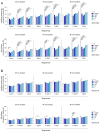
GPR84 agonists did not enhance J774-mediated phagocytosis of Raji cells. J774 macrophages, pre-treated with (A) 6-OAU or (B) ZQ-16, were incubated with LPS and co-cultured with pHrodo Red-labeled Raji cells and magrolimab for 6 h. Graphs show phagocytosis percentage and relative fluorescence intensity (RFI) of Raji cells at 2, 4, and 6 h. Data are normalized to macrophage numbers per well (n = 3 ± SEM). Statistical analysis: two-way ANOVA and Dunnett’s test, with * p < 0.05, ** p ≤ 0.01, and *** p ≤ 0.001.
Figure 5
Figure 5
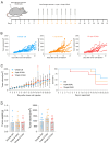
Oral 6-OAU administration showed no anti-cancer effects in the subcutaneous MC38 mouse model. C57BL/6 mice were injected with MC38 cells and treated with the vehicle or 6-OAU (1 or 10 mpk) via oral gavage. (A) The experimental design. (B) The tumor growth curve for each group. (C) Left: the average tumor growth and survival curve; right: the Kaplan–Meier curve. + indicates the removal of individual animals from experiments upon meeting termination criteria. (D) Tumor volume and weight at termination. Statistical analysis: (B,C) two-way ANOVA with Dunnett’s test; (D) one-way ANOVA with Tukey’s test.
Figure 6
Figure 6
Oral and intraperitoneal ZQ-16 administration showed no anti-cancer effects in the subcutaneous MC38 mouse model. C57BL/6 mice were injected with MC38 cells and treated with the vehicle or ZQ-16 (0.457 mpk IP or 10 mpk PO). (A) The experimental design. (B) The tumor growth curve for each group. (C) Left: the average tumor growth and survival curve; right: the Kaplan–Meier curve. + indicates the removal of individual animals from experiments upon meeting termination criteria. (D) Tumor volume and weight at termination. Statistical analysis: (B,C) two-way ANOVA with Dunnett’s test; (D) one-way ANOVA with Tukey’s test.

References

    1. Sawicki T., Ruszkowska M., Danielewicz A., Niedźwiedzka E., Arłukowicz T., Przybyłowicz K.E. A Review of Colorectal Cancer in Terms of Epidemiology, Risk Factors, Development, Symptoms and Diagnosis. Cancers. 2021;13:2025. doi: 10.3390/cancers13092025. - DOI - PMC - PubMed
    1. Kumar A., Gautam V., Sandhu A., Rawat K., Sharma A., Saha L. Current and emerging therapeutic approaches for colorectal cancer: A comprehensive review. World J. Gastrointest. Surg. 2023;15:495–519. doi: 10.4240/wjgs.v15.i4.495. - DOI - PMC - PubMed
    1. Pyo J., Park H.J. Treatment Efficacy of Immune Checkpoint Inhibitors for Patients with Advanced or Metastatic Colorectal Cancer: A Systematic Review and Meta-Analysis. J. Clin. Med. 2021;10:3599. doi: 10.3390/jcm10163599. - DOI - PMC - PubMed
    1. Borelli B., Antoniotti C., Carullo M., Germani M.M., Conca V., Masi G. Immune-Checkpoint Inhibitors (ICIs) in Metastatic Colorectal Cancer (mCRC) Patients beyond Microsatellite Instability. Cancers. 2022;14:4974. doi: 10.3390/cancers14204974. - DOI - PMC - PubMed
    1. Mantovani A., Allavena P., Marchesi F., Garlanda C. Macrophages as tools and targets in cancer therapy. Nat. Rev. Drug Discov. 2022;21:799–820. doi: 10.1038/s41573-022-00520-5. - DOI - PMC - PubMed

MeSH terms

Substances

LinkOut - more resources