. 2025 Jan 20;26(2):832.
doi: 10.3390/ijms26020832.

Cell Wall Microdomains Analysis in the Quadrifids of Utricularia dichotoma

Affiliations

Cell Wall Microdomains Analysis in the Quadrifids of Utricularia dichotoma

Bartosz J Płachno et al. Int J Mol Sci. .

Abstract

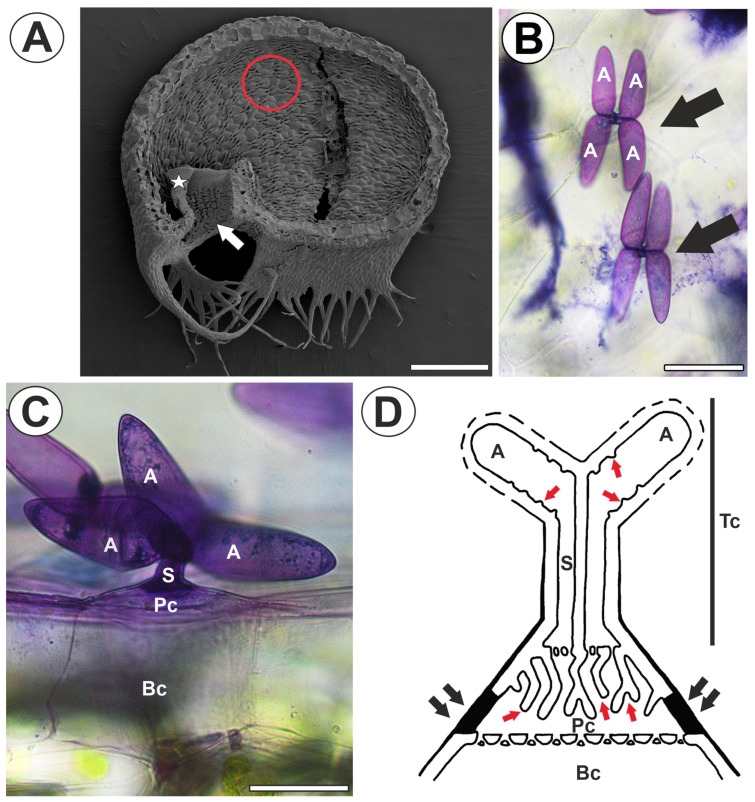
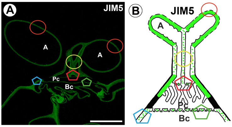
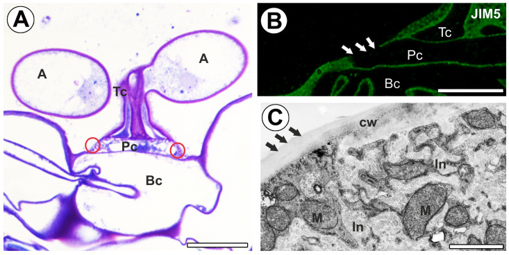
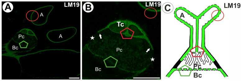
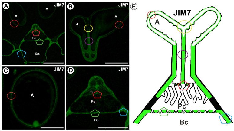
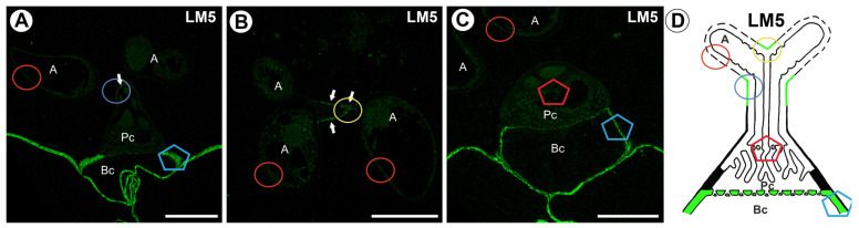
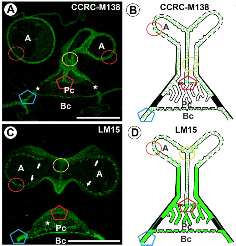
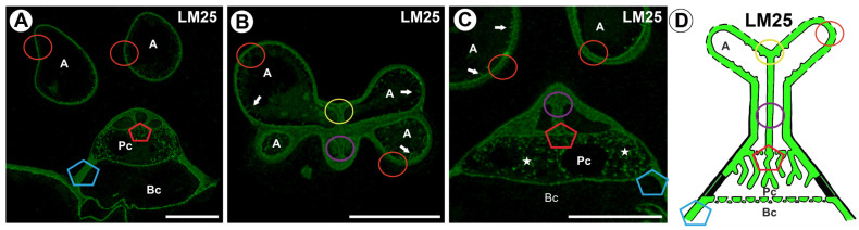
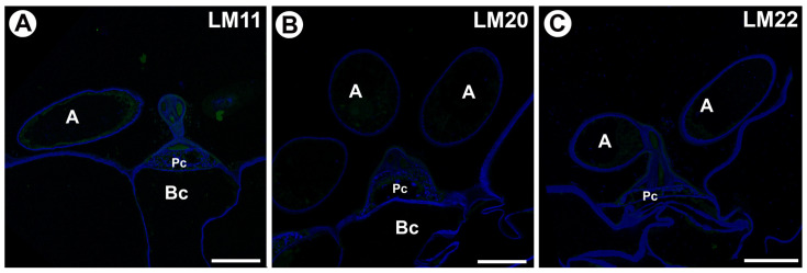
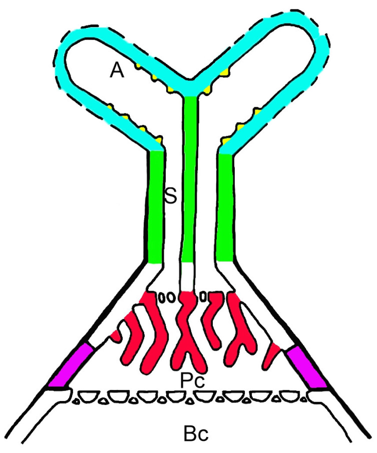
Carnivorous plants have fascinated botanists and ecologists with their various unusual adaptations in organ structure, physiology, and complex interactions with other organisms since the time of Charles Darwin. Species of the genus Utricularia (bladderworts, family Lentibulariaceae) are carnivorous plants that prey mainly on invertebrates using traps (bladders) of leaf origin. In the traps, there are glandular trichomes called quadrifids, which produce digestive enzymes and absorb the products of prey digestion. These quadrifids are unique due to their highly complex glandular cell structure; hence, they are an excellent model for studying the cell wall and its specialization. The main aim of the study was to investigate the presence and distribution of homogalacturonans (HGs) and hemicelluloses in the cell walls of trichome cells and especially in cell wall ingrowths in the quadrifid cells. The following antibodies were used against the wall components: anti-HGs (homogalacturonans) -JIM5 (low methylesterified HGs), JIM7 (highly esterified HGs), LM19 (low methylesterified HGs), CCRC-M38 (a fully de-esterified HG), LM5 (galactan); anti-hemicelluloses-LM25 (galactoxyloglucan; XXLLG, XXLG, XXXG modules of xyloglucans), LM15 (xyloglucan), CCRC-M138 (xylan), LM11 (heteroxylan); and anti-mannans: LM20 (heteromannan) and LM22 (heteromannan). The localization of the examined compounds was determined using immunohistochemistry techniques and immunogold labeling. In quadrifid cells, we found differences in the presence of the epitope detected by the LM5 antibody in the cell walls. In addition, cell wall ingrowths represented distinct microdomains of the cell wall in terms of the occurrence of wall components (they were methylesterified and demethylesterified homogalacturonan-poor). Hemicelluloses (galactoxyloglucan and xyloglucan) and arabinogalactans co-occur in cell wall ingrowths. Also, a part of the cell wall of the pedestal cell, which forms a Casparian strip, represented a distinct microdomain. We did not detect epitopes recognized by LM11, LM20 and LM22 antibodies. Our research shows that several cell wall microdomains occur in the cell walls of quadrifid cells. They differ depending on the presence and distribution of low methylesterified HGs, highly esterified HGs, fully de-esterified HGs, galactan (the epitope detected by the LM5 antibody), xyloglucan, galactoxyloglucan, and xylan (the epitope detected by the CCRC-M138 antibody).

Keywords: Lentibulariaceae; cell wall; cell wall microdomains; digestive trichomes; glands; hemicelluloses; pectic homogalacturonan; scanning transmission electron microscopy; transfer cells; xylan; xyloglucan.


Conflict of interest statement

The authors declare no conflicts of interest.

Figures

Figure 1
Figure 1
Trap structure of Utricularia dichotoma subsp. novae-zelandiae. Quadrifid structure. (A) A sagittally halved trap in scanning electron microscopy; trap door (star), trap entrance (arrow), with a red circle indicating the quadrifids, which are shown enlarged in (B), bar 500 µm. (B) The morphology of the quadrifids (arrow), treated with toluidine blue, arm (A), bar 20 µm. (C) Lateral view of the quadrifids, treated with toluidine blue; arm (A), stalk (s), pedestal cell (Pc), basal cell (Bc), bar 25 µm. (D) Diagram of quadrifid structure (after Fineran 1985 [40] modified); terminal cell (Tc), arm (A), stalk (s), pedestal cell (Pc), basal cell (Bc), cell wall ingrowths (red arrows), fully cutinized part of the cell wall of pedestal cell (black arrows), dashed line in arms indicates discontinuities in the cuticle.
Figure 2
Figure 2
Labeling of cells with JIM5 (low methylesterified HG) in the quadrifid (green color—signal of antibody). The section positions are shown with circles and polygons on a schematic representation of quadrifids. (A) Section through quadrifid: arm (A), pedestal cell (Pc), basal cell (Bc), bar 10 µm and bar 10 µm. (B) Diagram of occurrence of low methylesterified HGs (detected by JIM5) in quadrifid cells; the arm of the terminal cell (A), pedestal cell (Pc), basal cell (Bc).
Figure 3
Figure 3
Labeling of cells with JIM5 (low methylesterified HG) in the quadrifid (green color—signal of antibody). (A) Semi-thin section through quadrifid, arm (A), terminal cell (Tc), pedestal cell (Pc), fully cutinized part of the cell wall of pedestal cell (indicated with a red circle), basal cell (Bc), bar 10 µm. (B) Section through quadrifid (magnification of Figure 2A), terminal cell (Tc), pedestal cell (Pc), basal cell (Bc); note no positive signal in the fully cutinized part of the cell wall of pedestal cell (white arrows); bar 5 µm. (C) Part of the pedestal cell seen in scanning transmission electron microscopy, fully cutinized part of the cell wall of pedestal cell (black arrows), cell wall ingrowths (In), mitochondrion (M), cell wall (cw), bar 1 µm.
Figure 4
Figure 4
Labeling of cells with LM19 (low methylesterified HG) in the quadrifids (green color—a signal of antibody). The section positions are shown with circles and polygons on a schematic representation of quadrifids. (A,B) Section through quadrifid labeled with LM19, arm (A), terminal cell (Tc), pedestal cell (Pc), fully cutinized part of cell wall of pedestal cell (star), basal cell (Bc), wall ingrowths with positive signal (arrow); note part of pedestal cell without labeling (star); bar 10 µm and bar 10 µm. (C) Diagram of occurrence of low methylesterified HGs (detected by LM19) in quadrifid cells: the arm of the terminal cell (A), pedestal cell (Pc), basal cell (Bc).
Figure 5
Figure 5
Labeling of cells with CCRC-M38 (low methylesterified HG) in the quadrifids (green color—a signal of antibody). The section positions are shown with circles and polygons on a schematic representation of quadrifids. (A) Section through quadrifid labeled with CCRC-M38, arm (A), pedestal cell (Pc), basal cell (Bc), bar 10 µm. (B) Section through arms (A) labeled with CCRC-M38, bar 10 µm. (C) Section through the stalk of quadrifid, labeled with CCRC-M38, note four protoplasts (P), bar 10 µm. (D) Section through quadrifid labeled with CCRC-M38, terminal cell (TC), pedestal cell (Pc), basal cell (Bc), wall ingrowths with positive signal (arrow), bar 10 µm. (E) Diagram of occurrence of low methylesterified HGs (detected by CCRC-M38) in quadrifid cells: the arm of the terminal cell (A), pedestal cell (Pc), basal cell (Bc).
Figure 6
Figure 6
Labeling of cells with JIM7 (methylesterified HG) in the quadrifid (green color—signal of antibody). The section positions are shown with circles and polygons on a schematic representation of quadrifids. (A) Section through quadrifid, arm (A), pedestal cell (Pc), basal cell (Bc), bar 10 µm. (B) Section through arms (A), bar 10 µm. (C) Section through the arm (A), bar 10 µm. (D) Section through quadrifid, terminal cell (Tc), pedestal cell (Pc), basal cell (Bc) (magnification of Figure 6A), bar 10 µm. (E) Diagram of occurrence of methylesterified HGs (detected by JIM7) in quadrifid cells: the arm of the terminal cell (A), pedestal cell (Pc), basal cell (Bc).
Figure 7
Figure 7
Labeling of cells with LM5 (galactan) in the quadrifid (green color—signal of antibody). The section positions are shown with circles and polygons on a schematic representation of quadrifids. (A) Section through quadrifid, arm (A), pedestal cell (Pc), basal cell (Bc), positive signal in stalk indicated by arrow, bar 10 µm. (B) Section through arms (A), positive signal indicated by arrow, bar 10 µm. (C) Section through quadrifid, arm (A), pedestal cell (Pc), basal cell (Bc), bar 10 µm. (D) Diagram showing the occurrence of galactan (detected by LM5) in quadrifid cells: arm of terminal cell (A), pedestal cell (Pc), basal cell (Bc).
Figure 8
Figure 8
Labeling of cells with CCRC-M138 (xylan) and LM15 (xyloglucan) in the quadrifids (green color—a signal of antibody). The section positions are shown with circles and polygons on a schematic representation of quadrifids. (A) Section through quadrifid labeled with CCRC-M138, arm (A), pedestal cell (Pc), basal cell (Bc), note part of pedestal cell without labeling (star), bar 10 µm. (B) Diagram showing the occurrence of xylan (detected by CCRC-M138) in quadrifid cells: arm of terminal cell (A), pedestal cell (Pc), basal cell (Bc). (C) Section through quadrifid labeled with LM15, arm (A), pedestal cell (Pc), basal cell (Bc), positive signal in cell wall ingrowths in arms (arrow), note positive signal in cell wall ingrowths in pedestal cell (star), bar 10 µm. (D) Diagram showing the occurrence of xyloglucan (detected by LM15) in quadrifid cells: arm of terminal cell (A), pedestal cell (Pc), basal cell (Bc).
Figure 9
Figure 9
Labeling of cells with LM25 (galactoxyloglucans) in the quadrifids (green color—a signal of antibody). The section positions are shown with circles and polygons on a schematic representation of quadrifids. (A) Section through quadrifid, arm (A), pedestal cell (Pc), basal cell (Bc), bar 10 µm. (B) Section through arm of terminal cell (A), positive signal in cell wall ingrowths in arms (arrow), bar 10 µm (C) Section through quadrifid labeled, arm (A), pedestal cell (Pc), basal cell (Bc), positive signal in cell wall ingrowths in arms (arrow), note positive signal in cell wall ingrowths in pedestal cell (star), bar 10 µm. (D) Diagram showing the occurrence of galactoxyloglucans (detected by LM25) in quadrifid cells: arm of terminal cell (A), pedestal cell (Pc), basal cell (Bc).
Figure 10
Figure 10
Labeling of cells with LM11 (heteroxylan), LM20 (heteromannan) and LM22 (heteromannan) in the quadrifids (green color —a signal of antibody, blue color—cellulose stained by Calcofluor White), arm (A), pedestal cell (Pc), basal cell (Bc). (A) Section through quadrifid labeled with LM11, bar 10 µm. (B) Section through quadrifid labeled with LM20, bar 10 µm. (C) Section through quadrifid labeled with LM22, bar 10 µm.
Figure 11
Figure 11
A model depicting cell wall microdomains of quadrifids; Cell wall microdomains: cell wall ingrowths of the pedestal cell (red color), cell wall ingrowths of the terminal cell (yellow color), the cell wall of the stalk of the terminal cell (green color), the cell wall of the arm (blue color), and part of the cell wall of the pedestal cell (pink); basal cell (Bc), pedestal cell (Pc), stalk (S), arm (A).

Similar articles

References

    1. Juniper B.E., Robbins R.J., Joel D.M. The Carnivorous Plants. Academic Press; London, UK: 1989.
    1. Król E., Płachno B.J., Adamec L., Stolarz M., Dziubińska H., Trębacz K. Quite a few reasons for calling carnivores ‘the most wonderful plants in the world’. Ann. Bot. 2012;109:47–64. doi: 10.1093/aob/mcr249. - DOI - PMC - PubMed
    1. Ellison A.M., Adamec L. Carnivorous Plants: Physiology, Ecology, and Evolution. Oxford University Press; Oxford, UK: 2018. 510p
    1. Darwin C. Insectivorous Plants. 1st ed. John Murray; London, UK: 1875.
    1. Lloyd F.E. The range of structural and functional variety in the traps of Utricularia and Polypompholyx. Flora. 1932;126:303–328. doi: 10.1016/S0367-1615(17)31166-7. - DOI

LinkOut - more resources