Review
. 2025 Jan 20;26(2):846.
doi: 10.3390/ijms26020846.

De Novo DNM1L Pathogenic Variant Associated with Lethal Encephalocardiomyopathy-Case Report and Literature Review

Affiliations
Review

De Novo DNM1L Pathogenic Variant Associated with Lethal Encephalocardiomyopathy-Case Report and Literature Review

Martina Magistrati et al. Int J Mol Sci. .

Abstract

Pathogenic variants in DNM1L, encoding dynamin-like protein-1 (DRP1), cause a lethal encephalopathy. DRP1 defective function results in altered mitochondrial networks, characterized by elongated/spaghetti-like, highly interconnected mitochondria. We validated in yeast the pathogenicity of a de novo DNM1L variant identified by whole exome sequencing performed more than 10 years after the patient's death. Meanwhile, we reviewed the broadness and specificities of DNM1L-related phenotype. The patient, who exhibited developmental delay in her third year, developed a therapy-refractory myoclonic status epilepticus, followed by neurological deterioration with brain atrophy and refractory epilepsy. She died of heart failure due to hypertrophic cardiomyopathy. She was found to be heterozygous for the DNM1L variant (NM_ 012062.5):c.1201G>A, p.(Gly401Ser). We demonstrated its deleterious impact and dominant negative effect by assessing haploid and diploid mutant yeast strains, oxidative growth, oxygen consumption, frequency of petite, and architecture of the mitochondrial network. Structural modeling of p.(Gly401Ser) predicted the interference of the mutant protein in the self-oligomerization of the DRP1 active complex. DNM1L-related phenotypes include static or (early) lethal encephalopathy and neurodevelopmental disorders. In addition, there may be ophthalmological impairment, peripheral neuropathy, ataxia, dystonia, spasticity, myoclonus, and myopathy. The clinical presentations vary depending on mutations in different DRP1 domains. Few pathogenic variants, the p.(Gly401Ser) included, cause an encephalocardiomyopathy with refractory status epilepticus.

Keywords: DNM1L; burst suppression; global developmental regression; hypertrophic cardiomyopathy (HC); lethal encephalocardiomyopathy; refractory status epilepticus (RSE); retrospective post-mortem diagnosis.


Conflict of interest statement

All the authors report no conflicts of interest relevant to the manuscript.

Figures
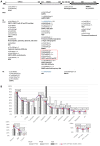
Figure 3
Figure 3
Genotype-phenotype correlation of DNM1L-related neurological disorders. (A) Pathogenic variants were distributed at each protein domain and according to their inheritance. Phenotypes due to each pathogenic variant are shown. EMPF1, encephalopathy due to defective mitochondrial and peroxisomal fission 1; OPA5, optic atrophy 5; HC, hypertrophic cardiomyopathy; MD, mitochondrial disease; NDD, neurodevelopmental disorder; RSE, Refractory Status Epilepticus; PR, paroxysmal refractory; IEM, inborn errors of metabolism; VD, variable domain; GED, GTPase effector domain; AD, autosomal dominant; AR, autosomal recessive; MOS, mosaic; §, in cis identified variants; × n, number of times a pathogenic variant has been described in unrelated patients (recurrent pathogenic variant); arrow, mutational hot spot; pathogenic variants with different inheritance patterns were underlined or colored in blue; in bold, the pathogenic variant identified in the present patient; †, variants for which lethality has been reported; in the dashed red box those pathogenic variants associated with epileptic encephalocardiomyopathy. Patient with isolated paroxysmal hemiparesis was reported by Zhang et al. [43]. (B) Distribution of the clinical features shown by DNM1L heterozygotes, including the present case, across the three protein domains, GTPase (white rectangles), MD (rectangles with dots) and GED (rectangles with diagonals). DD, developmental delay; E, encephalopathy; D Regr, Developmental Regression; C atrophy, cerebral atrophy; dysm/other, dysmyelination/Other brain RMN abnormalities; ophtalm, the presence of optic nerve atrophy and/or Poor visual fixation and/or nystagmus, and/or strabismus; RSE, refractory status epilepticus; S burst, suppression burst; Lactate, increased serum and/or CSF lactate; RCC skin, reduction of the activity of the respiratory chain complexes in skin; RCC muscle, reduction of the activity of the respiratory chain complexes in muscle. Percentages refer to the number of patients showing the clinical feature independently from the genotype subgroup. Among the numerous neurological features placed in order of decreasing frequency, those with impairment of higher brain functions have been grouped on the left. The biochemical items and those concerning epilepsy have been set apart.
Figure 1
Figure 1
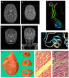
Brain MRI images showing rapidly progressive and diffuse cortical and subcortical atrophy, post-mortem presentation and histopathology of the heart, and structural consequences of the DRP1 p.(Gly401Ser) variant. (A) TSE T2 axial scans at the admission to the intensive care unit (at the age of 26 months) were comparable to those carried out at the age of 16 months. (B) TSE T2 axial scan on the same plane as the image shown in A was performed one month later (at 27 months). Brain MRI images show less cerebellar involvement compared to supratentorial structures, as shown in panel (B). (C) TSE T2 sagittal scan at the beginning of hospitalization (26 months) and (D) at the following month. In the protein 3D modeling for the DNM1L-wild-type (E), the white circle indicates the localization of 401 residues. The region of the Gly401Ser substitution (F): the wild-type protein is displayed in light blue, the mutated one in beige. Autoptic heart (G): weight was 110 g (proband’s weight 14.4 kg, at 61st centile). Coronal sections (H). Histology (20× panel (I), 40× panel (J)) with hematoxylin-eosin staining shows conspicuous myocellular hypertrophy and extensive interstitial fibrosis.
Figure 2
Figure 2
Functional analyses in the yeast S. cerevisiae. (A,B) Characterization of the haploid dnm1Δ strains harboring wild type allele (DNM1), the empty vector (dnm1Δ) or mutant allele dnm1G436S (G436S). (A) Oxidative growth: the strains were serially diluted and spotted on SC agar plates supplemented with the fermentable carbon source glucose (2%) or the non-fermentable carbon source K-acetate (2%) and incubated at 37 °C. (B) Mitochondrial morphology. For each strain, the percentage of the following morphotypes is reported: filamentous (in dark grey), network-like (in light grey) and linear (in black). (C,D) Characterization of the diploid DNM1/dnm1Δ strain harboring wild-type allele (DNM1/DNM1) mutant allele dnm1G436S (DNM1/G436S) or the empty vector (DNM1/dnm1Δ). (C) Oxidative growth: the strains were serially diluted and spotted on SC agar plates supplemented with the fermentable carbon source glucose (2%) or the non-fermentable carbon source K-acetate (2%) and incubated at 37 °C. (D) Mitochondrial morphology. For each strain, the percentage of the following morphotypes is reported: filamentous (in dark grey), network-like (in light grey) and linear (in black).

References

    1. Nasca A., Legati A., Baruffini E., Nolli C., Moroni I., Ardissone A., Goffrini P., Ghezzi D. Biallelic Mutations in DNM1L Are Associated with a Slowly Progressive Infantile Encephalopathy. Hum. Mutat. 2016;37:898–903. doi: 10.1002/humu.23033. - DOI - PMC - PubMed
    1. Yoon G., Malam Z., Paton T., Marshall C.R., Hyatt E., Ivakine Z., Scherer S.W., Lee K.-S., Hawkins C., Cohn R.D., et al. Lethal Disorder of Mitochondrial Fission Caused by Mutations in DNM1L. J. Pediatr. 2016;171:313–316.e1-2. doi: 10.1016/j.jpeds.2015.12.060. - DOI - PubMed
    1. Hogarth K.A., Costford S.R., Yoon G., Sondheimer N., Maynes J.T. DNM1L Variant Alters Baseline Mitochondrial Function and Response to Stress in a Patient with Severe Neurological Dysfunction. Biochem. Genet. 2018;56:56–77. doi: 10.1007/s10528-017-9829-2. - DOI - PubMed
    1. Barbet F., Hakiki S., Orssaud C., Gerber S., Perrault I., Hanein S., Ducroq D., Dufier J.-L., Munnich A., Kaplan J., et al. A Third Locus for Dominant Optic Atrophy on Chromosome 22q. J. Med. Genet. 2005;42:e1. doi: 10.1136/jmg.2004.025502. - DOI - PMC - PubMed
    1. Gerber S., Charif M., Chevrollier A., Chaumette T., Angebault C., Kane M.S., Paris A., Alban J., Quiles M., Delettre C., et al. Mutations in DNM1L, as in OPA1, Result in Dominant Optic Atrophy despite Opposite Effects on Mitochondrial Fusion and Fission. Brain. 2017;140:2586–2596. doi: 10.1093/brain/awx219. - DOI - PubMed