. 2025 Jan 8;14(1):45.
doi: 10.3390/pathogens14010045.

Characterization of Esocid Herpesvirus 1 (EsHV1) from Europe

Affiliations

Characterization of Esocid Herpesvirus 1 (EsHV1) from Europe

Mikael Leijon et al. Pathogens. .

Abstract

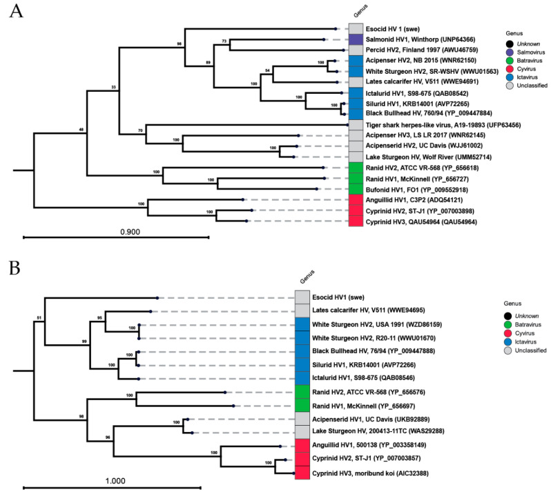
During routine sampling of northern pike, a male Esox lucius with circular blue-metallic granular spots mainly located on the head and back was identified. Histological investigations presented multifocally thickened epidermis rich in basophilic large structures with a granulated rim and a dense, non-granulated center. Other organs showed no signs of infection. Ultrastructural analysis of the skin revealed three different types of herpes-like structures predominantly located within enlarged vacuoles. PCR analysis and NGS of dissected skin tissue verified the presence of EsHV1 DNA. In this study, we describe the first identification of EsHV1 in mainland Europe. In addition, for the first time, full sequences of both the DNA polymerase and terminase of the virus is available, thus allowing for an improved phylogenetic placement of EsHV1 within the Alloherpesviridae family. In addition to the EsHV1 infected pike, we also observed that 11.1% of the pike were affected by lymphosarcoma, a hyperplasia-disease caused by retroviruses. In conclusion, viral infections in pike are relatively common and likely have consequences for the local population dynamics.

Keywords: Alloherpesviridae; EsHV1; Esox lucius; blue spot disease; genomic characterization; northern pike; pike.


Conflict of interest statement

The authors declare no conflict of interest.

Figures

Figure 1
Figure 1
EsHV1-infected pike with the characteristic blue spots. Zoomed-in view of the spots highlights the punctuated blue-metallic pattern.
Figure 2
Figure 2
Histological investigation of pike skin with epidermis and superficial layer of spongious dermis. (A) Epidermis of normal thickness, rich in mucous cells. (B) EsHV1-infected skin with thickened epidermis. The black square indicates the position of panel (C). (C) Dense with hypertrophied, herpesvirus-infected epidermal cells (arrow). (D) GaHV-1-infected epithelial cell (arrow) in cod gill for comparison.
Figure 3
Figure 3
Histological investigation of internal organs. (A) Liver with fully vacuolated cells with mild nuclear condensation. (B) Kidney, normal nephron including glomerulus (#), tubuli (¤), and the interstitium (*). A melanomachrophage ($) center can be seen. (C) Spleen, normal appearance with low immune activity. (D) Heart at the base/ventriculobulbar junction. V, ventricle; B, bulbus; VB, ventriculo-bulbar valves. Overlapping image: small focus of myocarditis in the spongiosum.
Figure 4
Figure 4
TEM analysis of infected skin. (A) Empty shells (A-capsids, red arrow) and mature capsids with clear centers (B-capsids, white arrow). (B) Mature C-capsids in large (black arrow), merged vacuoles (red arrow). (C) Higher-magnification image of mature C-capsids in close proximity to the Golgi apparatus and rough ER (black arrow).
Figure 5
Figure 5
Verification of EsHV1 using PCR. (A) Amplification of the terminase of exon 2 (TERM). (B) Amplification of the region of the EsHV1 polymerase on terminase exon 1 (POLIN-TERM). In (A), a 100-base-pair marker was used for reference, and in (B), a 1-kilo base pair marker. Triangular shapes and numbers above the gel images indicate annealing temperatures increasing in 2 °C increments.
Figure 6
Figure 6
Maximum likelihood phylogeny of the EsHV1-swe: (A) ca. 950 amino acids of the DNA polymerase and (B) DNA packing terminase comparing related and previously characterized alloherpesviruses infecting fish and amphibians. The Swedish isolate is highlighted in red. The genera as currently defined are shown with different colors. Virus names and isolate names are given when available; otherwise, country and year of collection are shown. Accession numbers for included sequences are given within parentheses. The bootstrap values of 100 replicates are shown at the nodes.
Figure 7
Figure 7
The North American (EsHV-1_usa) and Swedish (EsHV-1_swe) Esocid herpesvirus 1 with accessions KX198667 and PQ650602 aligned together with the corresponding region of Salmonid Herpes 1 (accession OK337613). Coding region annotations are as given in GenBank.

References

    1. Riha M., Gjelland K.O., Ded V., Eloranta A.P., Rabaneda-Bueno R., Baktoft H., Vejrik L., Vejrikova I., Drastik V., Smejkal M., et al. Contrasting structural complexity differentiate hunting strategy in an ambush apex predator. Sci. Rep. 2021;11:17472. doi: 10.1038/s41598-021-96908-1. - DOI - PMC - PubMed
    1. Nilsson J., Flink H., Tibblin P. Predator-prey role reversal may impair the recovery of declining pike populations. J. Anim. Ecol. 2019;88:927–939. doi: 10.1111/1365-2656.12981. - DOI - PubMed
    1. Ljunggren L., Sandström A., Bergström U., Mattila J., Lappalainen A., Johansson G., Sundblad G., Casini M., Kaljuste O., Eriksson B.K. Recruitment failure of coastal predatory fish in the Baltic Sea coincident with an offshore ecosystem regime shift. ICES J. Mar. Sci. 2010;67:1587–1595. doi: 10.1093/icesjms/fsq109. - DOI
    1. Tibblin P., Bergström K., Flink H., Hall M., Berggren H., Nordahl O., Larsson P. Higher abundance of adult pike in Baltic Sea coastal areas adjacent to restored wetlands compared to reference bays. Hydrobiologia. 2023;850:2049–2060. doi: 10.1007/s10750-023-05216-4. - DOI
    1. FishBase. 2023. [(accessed on 1 November 2024)]. Available online: https://www.fishbase.se/summary/SpeciesSummary.php?ID=258&AT=pike.

LinkOut - more resources