. 2025 Jan 10;14(1):57.
doi: 10.3390/pathogens14010057.

Eicosapentaenoic Acid and Docosahexaenoic Acid as an Antimicrobial Agent in Orthopedics-An In Vitro Study About the Race for Surface

Affiliations

Eicosapentaenoic Acid and Docosahexaenoic Acid as an Antimicrobial Agent in Orthopedics-An In Vitro Study About the Race for Surface

Christopher Spiegel et al. Pathogens. .

Abstract

Background: The burden of prosthetic joint infection in combination with antibiotic-resistant bacterial strains is a rising dilemma for patients experiencing total joint replacements. Around 0.8-2% of patients experience prosthetic joint infections, while up to 21% of patients are considered fatal cases after 5 years. Staphylococcus aureus is one of the main reasons for prosthetic joint infections. Its capability of forming biofilms and developing mechanisms against antibiotics is one of the most dangerous clinical topics being currently discussed. Previous studies have shown the promising results of omega-3 fatty acids as an antimicrobial agent against Staphylococcus aureus. Though an antimicrobial effect has been examined, the influence of polyunsaturated fatty acids on Staphylococcus aureus in the presence of human osteoblasts has not been reported yet. In this study, we aimed to investigate the influence of omega-3 fatty acids on the biofilm formation of Staphylococcus aureus ATCC 29213 in the presence of hFOB 1.19 cells. The co-culture setup helped to examine the influence of omega-3 fatty acids on the race for surface to simulate prosthetic joint infections.

Methods: In this study, we tested Staphylococcus aureus ATCC 29213 co-cultured with human fetal osteoblasts hFOB 1.19 in the presence of sub-MIC and MIC concentrations of docosahexaenoic acid (1.25 mg/L, 2.5 mg/L) and eicosapentaenoic acid (0.15 mg/L, 0.3 mg/L) after 1, 6 and 24 h of incubation. After establishing the co-culture, cell culture and biofilm, we performed colony-forming unit counting and cell counting to examine cell survivability. In addition, we carried out scanning electron microscopy to study the race for surface behaviour of the cells.

Results: We found a protective influence of omega-3 fatty acids on osteoblasts when present in co-culture with Staphylococcus aureus after 6 h of incubation. Omega-3 fatty acids increase the cell survival of osteoblasts after 6 h in a co-culture with bacteria and are able to influence the race for surface. In this study, the strain of Staphylcoccus aureus ATCC 29213 showed signs of growth inhibition within the first 6 h.

Conclusions: Omega-3 fatty acids can be a valuable antimicrobial agent in terms of decreasing the risk of on-site infection during surgery. Omega-3 fatty acids were shown to decrease the bacterial load within the first 6 h of incubation and increase the survivability of osteoblasts.

Keywords: PJI; Staphylococcus aureus; biofilm; implant-related infections; omega-3 fatty acids; orthopedics.


Conflict of interest statement

The authors declare no conflicts of interest.

Figures

Figure 1
Figure 1
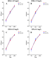
hFOB 1.19 osteoblast cell count under the influence of PUFAs after 1, 6 and 24 h of incubation. The results are plotted against matching solvent controls and controls to understand the influence of PUFAs against ethanol. The differences between each nutrient medium and its glucose alternative were analyzed using a two-way ANOVA followed by Dunnett’s multiple comparison tests. The results are presented as mean values ± SD, derived from three independent experiments conducted in triplicate.
Figure 2
Figure 2
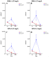
Colony-forming units of Staphylococcus aureus ATCC 29213 in co-culture with hFOB 1.19 osteoblasts under the influence of PUFAs after 1, 6 and 24 h of incubation. The results are plotted against matching ethanol concentration and controls to understand the influence of PUFAs against ethanol. The differences between each nutrient medium and its glucose alternative were analyzed using a two-way ANOVA followed by Dunnett’s multiple comparison tests. The results are presented as mean values ± SD, derived from three independent experiments conducted in triplicate.
Figure 3
Figure 3
Osteoblast cell count in a co-culture with Staphylococcus aureus ATCC 29213 under the influence of PUFAs after 1, 6 and 24 h of incubation. The results show matching ethanol concentration and controls to understand the activity of PUFAs in the presence of ethanol. For a proper presentation, absolute cell counts are shown in the figures. The differences between each nutrient medium and its glucose alternative were analyzed using a two-way ANOVA followed by Dunnett’s multiple comparison tests. The results are presented as mean values ± SD, derived from three independent experiments conducted in triplicate.
Figure 4
Figure 4
Control of osteoblast cell adhesion and growth after 1, 6 and 24 h in co-culture with Staphylococcus aureus ATCC 29213 at a PUFA concentration of 0 mg/L.
Figure 5
Figure 5
Osteoblast cell adhesion and growth after 1, 6 and 24 h in a co-culture with Staphylococcus aureus ATCC 29213 at a PUFA concentration of 1.25 mg/L DHA.
Figure 6
Figure 6
Osteoblast cell adhesion and growth after 1, 6 and 24 h in a co-culture with Staphylococcus aureus ATCC 29213 at a PUFA concentration of 2.5 mg/L DHA.
Figure 7
Figure 7
Osteoblast cell adhesion and growth after 1, 6 and 24 h in a co-culture with Staphylococcus aureus ATCC 29213 at a PUFA concentration of 0.15 mg/L EPA.
Figure 8
Figure 8
Osteoblast cell adhesion and growth after 1, 6 and 24 h in a co-culture with Staphylococcus aureus ATCC 29213 at a PUFA concentration of 0.3 mg/L EPA.

Similar articles

References

    1. Ahmed S.S., Begum F., Kayani B., Haddad F.S. Risk factors, diagnosis and management of prosthetic joint infection after total hip arthroplasty. Expert Rev. Med. Devices. 2019;16:1063–1070. doi: 10.1080/17434440.2019.1696673. - DOI - PubMed
    1. Jin X., Luxan B.G., Hanly M., Pratt N.L., Harris I., de Steiger R., Graves S.E., Jorm L. Estimating incidence rates of periprosthetic joint infection after hip and knee arthroplasty for osteoarthritis using linked registry and administrative health data. Bone Jt. J. 2022;104:1060–1066. doi: 10.1302/0301-620X.104B9.BJJ-2022-0116.R1. - DOI - PMC - PubMed
    1. Tao Y., Luo Y., Hu H., Wang W., Zhao Y., Wang S., Zheng Q., Zhang T., Zhang G., Li J., et al. Clinically applicable optimized periprosthetic joint infection diagnosis via AI based pathology. NPJ Digit. Med. 2024;7:303. doi: 10.1038/s41746-024-01301-7. - DOI - PMC - PubMed
    1. Natsuhara K.M., Shelton T.J., Meehan J.P., Lum Z.C. Mortality During Total Hip Periprosthetic Joint Infection. J. Arthroplast. 2019;34:S337–S342. doi: 10.1016/j.arth.2018.12.024. - DOI - PubMed
    1. Okike K., Bhattacharyya T. Trends in the management of open fractures. A critical analysis. JBJS. 2006;88:2739–2748. doi: 10.2106/00004623-200612000-00025. - DOI - PubMed

MeSH terms

LinkOut - more resources