. 2025 Jan 20;17(2):359.
doi: 10.3390/nu17020359.

Differential Enhancement of Fat-Soluble Vitamin Absorption and Bioefficacy via Micellization in Combination with Selected Plant Extracts In Vitro

Affiliations

Differential Enhancement of Fat-Soluble Vitamin Absorption and Bioefficacy via Micellization in Combination with Selected Plant Extracts In Vitro

Stefanie Steinbauer et al. Nutrients. .

Abstract

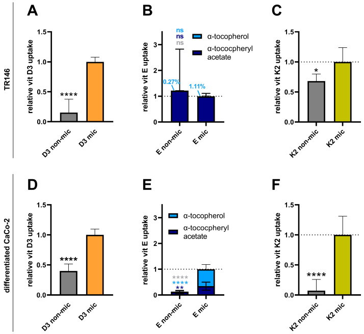
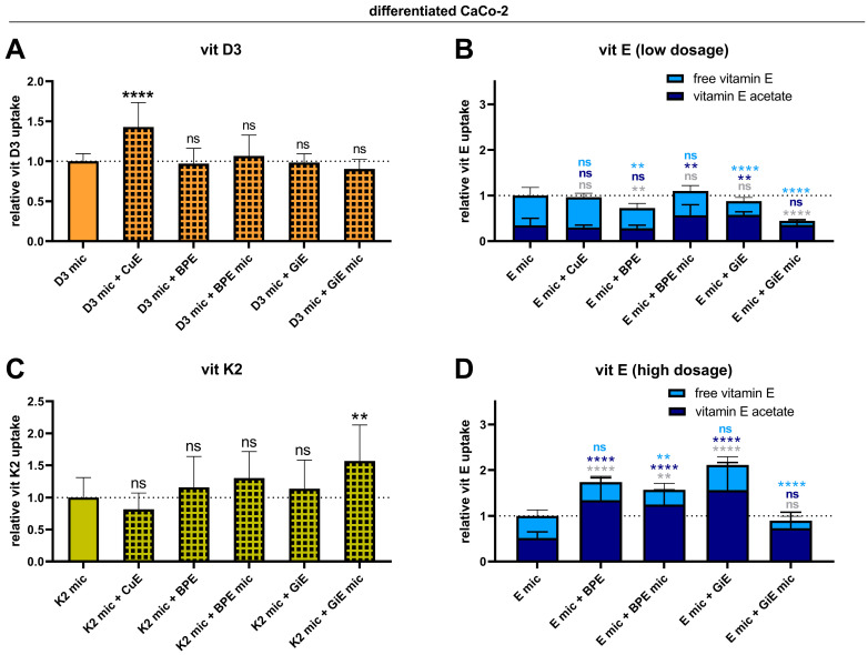
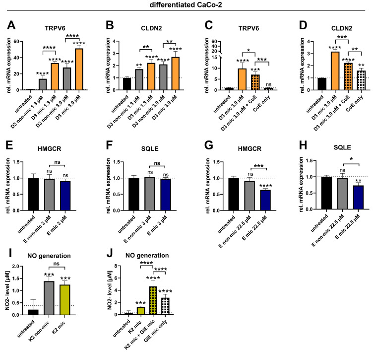
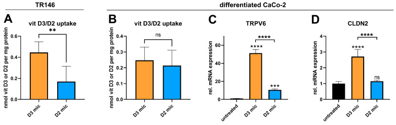
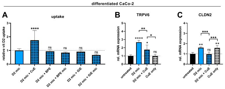
Background/Objectives: Individuals with special metabolic demands are at risk of deficiencies in fat-soluble vitamins, which can be counteracted via supplementation. Here, we tested the ability of micellization alone or in combination with selected natural plant extracts to increase the intestinal absorption and bioefficacy of fat-soluble vitamins. Methods: Micellated and nonmicellated vitamins D3 (cholecalciferol), D2 (ergocalciferol), E (alpha tocopheryl acetate), and K2 (menaquionone-7) were tested in intestinal Caco-2 or buccal TR146 cells in combination with curcuma (Curcuma longa), black pepper (Piper nigrum), or ginger (Zingiber officinale Roscoe) plant extracts. The vitamin uptake was quantified via HPLC-MS, and bioefficacy was assessed via gene expression analyses or the Griess assay for nitric oxide generation. Results: Micellization increased the uptake of vitamin D into buccal and intestinal cells, with vitamin D3 being more efficient than vitamin D2 in increasing the expression of genes involved in calcium transport. The micellization of vitamin E acetate increased its uptake and conversion into biologically active free vitamin E in intestinal cells only. The vitamin K2 uptake into buccal and intestinal cells was increased via micellization. Plant extracts increased the uptake of select micellated vitamins, with no plant extract being effective in combination with all vitamins. The curcuma extract increased the uptake of vitamins D2/D3 but not their bioefficacy. Black pepper and ginger extracts increased the uptake of vitamin E acetate into intestinal cells but failed to increase its conversion into free vitamin E. The ginger extract augmented the uptake of vitamin K2 and increased NO generation additively. Conclusions: Our data substantiate the positive effects of micellization on fat-soluble vitamin absorption and bioefficacy in vitro. While the application of plant extracts in addition to micellization to further increase bioefficacy is an interesting approach, further studies are warranted to understand vitamin-specific interactions and translation into increased bioefficacy.

Keywords: alpha tocopherol acetate; bioavailability; buccal absorption; cholecalciferol; ergocalciferol; intestinal absorption; intestinal bioefficacy; menaquinone-7; micellization; vitamin–plant–compound interactions.


Conflict of interest statement

Author Stefanie Steinbauer, Melanie Wallner, Theresa Gramatte, Julian Weghuber and Bernhard Blank-Landeshammer were employed by the company FFoQSI GmbH—Austrian Competence Centre for Feed and Food Quality, Safety and Innovation. M.I. is employed by PM International AG, which provided funding for the Austrian Competence Centre for Feed and Food Quality, Safety and Innovation and the Josef Ressel Center for Phytogenic Drug Research. PM International AG had no influence on the study design or the decision to report the research. The other authors declare no conflicts of interest.

Figures

Figure 1
Figure 1
Effect of micellization on the buccal and intestinal uptake of vitamins D3, E, and K2. TR146 (AC) or differentiated Caco-2 cells (DF) were treated with vitamins (vitamin D3, 3.9 µM; vitamin E acetate, 3 µM; vitamin K2, 2.3 µM) in either nonmicellated (“non-mic”) or micellated forms (“mic”) for 4 h. Afterward, vitamins were extracted and quantified via HPLC–MS analysis. The data were derived from two (A,CF) or four (B) independent experiments performed in triplicate or quadruplicate. (B): The Mann–Whitney test was used for the statistical analysis.* p ≤ 0.05, ** p ≤ 0.01 and **** p ≤ 0.0001. “ns” indicates no statistical significance. Significance levels in (B,E) refer to comparisons the levels of α-tocopheryl acetate (dark blue), α-tocopherol (light blue) and total vitamin E (grey).
Figure 2
Figure 2
Effects of micellization in combination with plant extracts on the intestinal uptake of vitamins D3, E, and K2. Differentiated Caco-2 cells were either treated with micellated vitamins (vitamin D3, 3.9 µM (A); vitamin E acetate, 3 µM (B) and 22.5 µM (D); or vitamin K2, 2.3 µM (C) alone or in combination with CuE (CuE, 14.7 µg/mL), nonmicellated BPE (BPE, 6 µg/mL), micellated BPE (BPE mic, 6 µg/mL), nonmicellated GiE (GiE, 58.8 µg/mL), or micellated GiE (GiE mic, 58.8 µg/mL) for 4 h. Afterward, vitamins were extracted and quantified via HPLC–MS analysis. The data shown were obtained from two to four independent experiments performed in triplicate. ** p ≤ 0.01, **** p ≤ 0.0001. “ns” indicates no statistical significance. Significance levels in (B,D) refer to comparisons the levels of α-tocopheryl acetate (dark blue), α-tocopherol (light blue) and total vitamin E (grey).
Figure 3
Figure 3
Effects of micellization alone or in combination with plant extracts on biological markers of vitamin D3, E, and K2 function in Caco-2 cells. Differentiated Caco-2 cells were either not treated with the test substances (“untreated”) or treated with the vitamins (vitamin D3, 1.3 µM, 3.9 µM; vitamin E acetate, 3 µM and 22.5 µM; vitamin K2, 2.3 µM) in the nonmicellated form (“non-mic”), micellated form (“mic”), or micellated form in combination with CuE (CuE, 14.7 µg/mL) or micellated GiE (GiE mic, 58.8 µg/mL) for 24 h. Afterward, RNA was extracted and analyzed for TRPV6, CLDN2, HMGCR, or SQLE expression via RT–qPCR (AH), or the nitrite concentration in the cell supernatant was measured using the Griess assay (I,J). The data shown were obtained from two independent experiments performed in duplicate (AH) or triplicate (I,J). * p ≤ 0.05, ** p ≤ 0.01, *** p ≤ 0.001 and **** p ≤ 0.0001. “ns” indicates no statistical significance.
Figure 4
Figure 4
Comparison of vitamin D3 and vitamin D2 in terms of their buccal and intestinal absorption efficiencies and effects on intestinal calcium absorption markers. TR146 (A) or differentiated Caco-2 cells (BD) were either untreated or treated with micellated vitamin D3 (D3 mic; 3.9 µM) or micellated vitamin D2 (D2 mic; 3.9 µM) for 4 h (A,B) or 24 h (C,D). After 4 h of treatment, vitamins D3 and D2 were extracted and quantified via HPLC–MS analysis. After 24 h of treatment, RNA was extracted and analyzed for TRPV6 or CLDN2 expression via RT–qPCR (C,D). The data were obtained from two independent experiments performed in quadruplicate (A), five independent experiments performed in triplicate (B), or two independent experiments performed in duplicate (C,D). ** p ≤ 0.01, *** p ≤ 0.001 and **** p ≤ 0.0001. “ns” indicates no statistical significance.
Figure 5
Figure 5
Effect of micellization in combination with plant extracts on intestinal vitamin D2 uptake and on biological markers of vitamin D function in Caco-2 cells. Differentiated Caco-2 cells were either untreated or treated with micellated vitamin D2 (D2 mic, 3.9 µM) alone or in combination with CuE (CuE, 14.7 µg/mL), nonmicellated BPE (BPE, 6 µg/mL), micellated BPE (BPE mic, 6 µg/mL), nonmicellated GiE (GiE, 58.8 µg/mL), or micellated GiE (GiE mic, 58.8 µg/mL) for 4 h (A) or with D2 mic and/or CuE for 24 h (B,C). After 4 h of treatment, vitamin D2 was extracted and quantified via HPLC–MS analysis. After 24 h of treatment, RNA was extracted and analyzed for TRPV6 and CLDN2 expression via RT–qPCR. The data shown were obtained from two to four independent experiments performed in triplicate (A) or two independent experiments performed in duplicate (B,C). * p ≤ 0.05, ** p ≤ 0.01, *** p ≤ 0.001 and **** p ≤ 0.0001. “ns” indicates no statistical significance.

Similar articles

References

    1. Fleet J.C. Vitamin D-Mediated Regulation of Intestinal Calcium Absorption. Nutrients. 2022;14:3351. doi: 10.3390/nu14163351. - DOI - PMC - PubMed
    1. Kumar M., Deshmukh P., Bhatt A., Sinha A.H., Chawla P. Vitamin E Supplementation and Cardiovascular Health: A Comprehensive Review. Cureus. 2023;15:e48142. doi: 10.7759/cureus.48142. - DOI - PMC - PubMed
    1. Lai Y., Masatoshi H., Ma Y., Guo Y., Zhang B. Role of Vitamin K in Intestinal Health. Front. Immunol. 2021;12:791565. doi: 10.3389/fimmu.2021.791565. - DOI - PMC - PubMed
    1. Desmarchelier C., Borel P., Goncalves A., Kopec R., Nowicki M., Morange S., Lesavre N., Portugal H., Reboul E. A Combination of Single-Nucleotide Polymorphisms Is Associated with Interindividual Variability in Cholecalciferol Bioavailability in Healthy Men. J. Nutr. 2016;146:2421–2428. doi: 10.3945/jn.116.237115. - DOI - PubMed
    1. Traber M.G., Sies H. Vitamin E in humans: Demand and delivery. Annu. Rev. Nutr. 1996;16:321–347. doi: 10.1146/annurev.nu.16.070196.001541. - DOI - PubMed

LinkOut - more resources