. 2024 Dec 30;17(1):34.
doi: 10.3390/pharmaceutics17010034.

Sulforaphane Wrapped in Self-Assembled Nanomicelle Enhances the Effect of Sonodynamic Therapy on Glioma

Affiliations

Sulforaphane Wrapped in Self-Assembled Nanomicelle Enhances the Effect of Sonodynamic Therapy on Glioma

Yihong Li et al. Pharmaceutics. .

Abstract

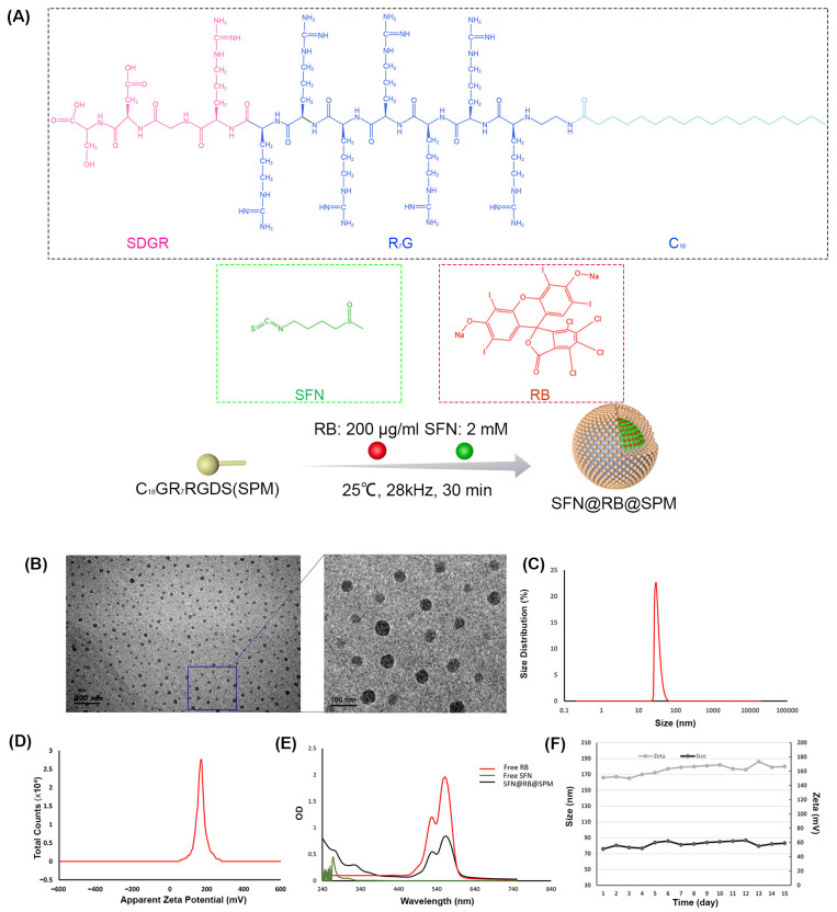
Background/Objectives: The two obstacles for treating glioma are the skull and the blood brain-barrier (BBB), the first of which forms a physical shield that increases the difficulties of traditional surgery or radiotherapy, while the latter prevents antitumor drugs reaching tumor sites. To conquer these issues, we take advantage of the high penetrating ability of sonodynamic therapy (SDT), combined with a novel nanocomplex that can easily pass the BBB. Methods: Through ultrasonic polymerization, the amphiphilic peptides (C18GR7RGDS) were self-assembled as a spherical shell encapsulating a sonosensitizer Rose Bengal (RB) and a plant-derived compound, sulforaphane (SFN), to form the nanocomplex SFN@RB@SPM. Results/Conclusions: SFN@RB@SPM can be internalized by the glioma cells through the tumor-targeting motif RGDS (abbreviated for the peptide sequence composed of arginine, glycine, aspartic acid, and serine), and further executes antitumor function during SDT. Also, SFN@RB@SPM could be easily taken up by U87-MG cells and cross the BBB in glioma-bearing mice during SDT. The mechanism investigation revealed that, compared with the SFN-free nanocomplex (RB@SPM), SFN@RB@SPM induced much more apoptosis of U87-MG cells in an ROS-dependent manner through the depletion of glutathione by SFN and the cavitation effect by SDT. In animal experiments, besides a significant reduction in tumor volume and a delay in losing body weight, H&E staining showed a massive infiltration of neutrophils adjacent to the tumor sites, indicating this novel nanocomplex SFN@RB@SPM can synergistically augment SDT efficacy, partially by enhancing the antitumor function of innate immunity.

Keywords: glioma; neutrophil infiltration; self-assembled nanomicelle; sonodynamic therapy; sulforaphane.


Conflict of interest statement

The authors declare that they have no known competing financial interests or personal relationships that could have appeared to influence the work reported in this paper.

Figures

Figure 1
Figure 1
Characterization of self-assembled nanocomplex SFN@RB@SPM. (A) Chemical structures of C18GR7RGDS (SPM), RB, and SFN. For the molecular composition of SPM, as illustrated, the pink hydrophilic sequence SDGR is conjugated to the green hydrophobic sequence C18 through the blue linker sequence R7G. At the reaction concentration of 200 μg/mL of RB and 2 mM of SFN, after being sonicated (25 °C, 28 kHz for 30 min) for polymerization, RB and SFN could be encapsulated into the self-assembled nanomicelle (SPM) to form a new nanocomplex, SFN@RB@SPM. (B) The nanocomplex SFN@RB@SPM was visualized by transmission electron microscope with 80,000× magnification. Scale bar, 200 nm for the left photo and 100 nm for the right one. (C) Size distribution indicates the mean size of SFN@RB@SPM is approximately 57 nm. (D) The apparent zeta potential of SFN@RB@SPM is 166 ± 0.81 mV. (E) UV–VIS absorption experiment manifested the specific peaks for SFN@RB@SPM, confirming that the free SFN and RB had been successfully enveloped into SPM. (F) The size and zeta potential were measured every day for 15 days, with the results indicating good water stability.
Figure 2
Figure 2
Cellular uptake of SFN@RB@SPM. U87-MG cells were treated with SFN@RB@SPM at the concentration of 10 μM (normalized to the molar concentration of SFN) for 30 min, 1 h, and 4 h, and then the internalization of SFN@RB@SPM (monitored by red fluorescence of RB) by U87-MG cells was visualized under a laser confocal fluorescence microscope. The nuclei were stained with DAPI (blue). Scale bar, 20 μm.
Figure 3
Figure 3
SFN@RB@SPM shows synergistic cytotoxicity with US irradiation in vitro. (A) The upper panel shows that U87-MG cells were treated with control, SFN (10 μM), RB@SPM (1 μM), or SFN@RB@SPM (normalized to the molar concentration of 10 μM SFN) with or without US irradiation for 24 h, where US (ultrasound irradiation) refers to high energy ultrasound waves exciting a sonosensitizer, resembling a process of light irradiation in PDT. In contrast, the lower panel shows that U87-MG cells were pretreated with control, SFN (10 μM), RB@SPM (1 μM) or SFN@RB@SPM (normalized to the molar concentration of 10 μM SFN) for 4 h and then exposed to US (1 MHz, 1 W/cm2) for 24 h. Then the cells were stained with Calcein-AM to show live (green) and dead (red) cells. Scale bar, 100 μm. (B) After the same treatment procedures as in (A), an MTT assay was conducted to determine cell viability. The data represent the mean ± SEM of three independent experiments. ** p < 0.01, *** p < 0.001 versus control. (C) U87-MG cells were subjected to flow cytometry to detect the apoptosis induced by SFN, RB@SPM, and SFN@RB@SPM with or without US irradiation (1 MHz, 1 W/cm2). The cells were double-stained with Annexin V/PI, and the apoptotic ratio was quantified in (D), expressed as mean ± SEM of three independent experiments. *** p < 0.001 versus control.
Figure 4
Figure 4
SFN@RB@SPM augments the cytotoxic effect of SDT in vitro in an ROS-dependent manner through the synergism of SFN-mediated GSH depletion and RB-mediated elevation of ROS. (A) U87-MG cells were pretreated with control, SFN (10 μM), RB@SPM (1 μM), or SFN@RB@SPM (normalized to the molar concentration of 10 μM SFN) for 4 h, with or without following US irradiation (1 MHz, 1 W/cm2) for 30 s. ROS levels were then measured in the cells using Dichlorofluorescein (DCF). As indicated, the intensity of green fluorescence is in proportion with the intracellular level of ROS. Scale bar, 50 μm. (B) The quantification of fluorescence intensity in (A). Data represent the mean ± SEM of three independent experiments. ** p < 0.01, *** p < 0.001 versus control. (C) U87-MG cells were treated with the same procedures in (A), and then the intracellular GSH contents were measured using a GSH detection kit. Data represent the mean ± SEM of three independent experiments. * p < 0.05, ** p < 0.01 versus control. (D) U87-MG cells were pretreated with or without N-acetylcysteine (NAC), and then these two groups were treated in the same manner as in (A), and the cell viability was determined 24 h later using CCK8 assay. Data represent the mean ± SEM of three independent experiments. * p < 0.05, *** p < 0.001 versus control.
Figure 5
Figure 5
SFN@RB@SPM exhibits a robust antitumor activity for the glioma xenograft of nude mice in coordination with SDT. (A) Schematic illustration showing the time window and the route of drug administration in combination with SDT for the glioma-bearing nude mice. Mice were intravenously injected with PBS, RB, SFN, RB@SPM, or SFN@RB@SPM, with or without following US irradiation every 3 days for five repetitions of the treatments. The mice were then sacrificed at day 20. (B) Glioma-bearing mice were randomly divided into six groups (n = 6) treated with PBS, PBS plus US, RB plus US, SFN plus US, RB@SPM plus US, and SFN@RB@SFN plus US. The bioluminescence was recorded by the IVIS Spectrum imaging system at different time points (1 d, 4 d, 7 d, 10 d, 13 d, 20 d) post treatments. (C) The tumor volume in each group of mice was determined using IVIS Spectrum, and the data represent the mean volume of tumor ± SEM in each group of mice. ** p < 0.01 versus control. (D) The body weight was recorded at the indicated time points, and the data represent the mean ± SEM in each group of mice. ** p < 0.01 versus control. (E) The mice of the above six groups were sacrificed at day 20, and the brain tissues were isolated for TUNEL (the first panel) and H&E staining (the last two panels). The scale bars of photos from first to third panel are 100 μm, 100 μm and 50 μm, respectively. The green fluorescence represents the apoptotic cells. The middle panel displays images of lower magnification (20×) for H&E staining. The green dotted lines within the images indicate the boundary between the tumor and normal tissues. In the SFN@RB@SPM plus US-treated group of the third panel, a large number of neutrophils have infiltrated (indicated by the arrowheads) into the area adjacent to the tumor tissue. This rectangular area of the 40× image is further magnified.
Figure 6
Figure 6
A diagram illustrating the potential mechanism by which SFN@RB@SPM synergistically improves the efficacy of traditional SDT. SFN and RB are encapsulated into a self-assembled nanomicelle (C18GR7RGDS) under ultrasound polymerization. The orthotopic glioma-bearing nude mice were intravenously injected with SFN@RB@SPM, and after 2 h, the mice were treated with SDT. The RGDS-motif guides the nanocomplex to cross the BBB and further penetrate the tumor tissues through SDT-induced transient BBB-opening. While approaching the tumor cells that express the specific receptor for the RGDS-motif, SFN@RB@SPM is internalized and releases the payload comprising SFN and RB. RB may directly promote the generation of ROS by an SDT-induced cavitation effect like the pyrolysis of water within the tumor cells. In addition, the released SFN can react with GSH or its precursor NAC to form SFN-GSH or SFN-NAC, both of which can significantly diminish the GSH level. Furthermore, the instant BBB opening and the massive numbers of ROS may activate neutrophils, which further execute the antitumor function and synergistically augment the SDT effect.

Similar articles

Cited by

References

    1. Gilbert M.R., Dignam J.J., Armstrong T.S., Wefel J.S., Blumenthal D.T., Vogelbaum M.A., Colman H., Chakravarti A., Pugh S., Won M., et al. A randomized trial of bevacizumab for newly diagnosed glioblastoma. N. Engl. J. Med. 2014;370:699–708. doi: 10.1056/NEJMoa1308573. - DOI - PMC - PubMed
    1. Chinot O.L., Wick W., Mason W., Henriksson R., Saran F., Nishikawa R., Carpentier A.F., Hoang-Xuan K., Kavan P., Cernea D., et al. Bevacizumab plus radiotherapy-temozolomide for newly diagnosed glioblastoma. N. Engl. J. Med. 2014;370:709–722. doi: 10.1056/NEJMoa1308345. - DOI - PubMed
    1. Bucci M.K., Maity A., Janss A.J., Belasco J.B., Fisher M.J., Tochner Z.A., Rorke L., Sutton L.N., Phillips P.C., Shu H.K. Near complete surgical resection predicts a favorable outcome in pediatric patients with nonbrainstem, malignant gliomas: Results from a single center in the magnetic resonance imaging era. Cancer. 2004;101:817–824. doi: 10.1002/cncr.20422. - DOI - PubMed
    1. Fan W., Yung B., Huang P., Chen X. Nanotechnology for Multimodal Synergistic Cancer Therapy. Chem. Rev. 2017;117:13566–13638. doi: 10.1021/acs.chemrev.7b00258. - DOI - PubMed
    1. Eichler A.F., Chung E., Kodack D.P., Loeffler J.S., Fukumura D., Jain R.K. The biology of brain metastases-translation to new therapies. Nat. Rev. Clin. Oncol. 2011;8:344–356. doi: 10.1038/nrclinonc.2011.58. - DOI - PMC - PubMed

LinkOut - more resources