. 2025 Jan 7;17(1):73.
doi: 10.3390/pharmaceutics17010073.

Retinal Protection of New Nutraceutical Formulation

Affiliations

Retinal Protection of New Nutraceutical Formulation

Luca Rosario La Rosa et al. Pharmaceutics. .

Abstract

Background/Objectives: Retinal ganglion cell (RGC) protection represents an unmet need in glaucoma. This study assessed the neuroprotective, antioxidant, and anti-inflammatory effect of a new nutraceutical formulation named Epicolin, based on citicoline, homotaurine, epigallocatechin-3-gallate, forskolin, and vitamins, through in vitro and in vivo studies. Methods: The neuroprotective effect of Epicolin or its single components, and Epicolin compared to an untreated control and two marketed formulations [Formulation G (FG) and N (FN)], was evaluated in neuroblastoma cells (SH-SY5Y) challenged with staurosporine. The antioxidant potential and the scavenging activity of Epicolin compared to the untreated control, and FG and FN, was evaluated in SH-SY5Y cells and through oxygen radical absorbance capacity acellular assay, respectively. Moreover, the protective effect against hypoxic damage was evaluated in Muller cells (MIO-M1) subjected to hypoxia. The efficacy of Epicolin was also evaluated in DBA/2J glaucomatous mice through the use of a pattern electroretinogram (PERG), immunostaining, and real-time PCR. Results: Among the nutraceutical formulations tested, only Epicolin showed a significant neuroprotective effect on SH-SY5Y attributable to the synergistic action of its single ingredients. As for antioxidant and scavenging activity, Epicolin showed a higher efficacy compared to FG and FN. Furthermore, Epicolin showed the same protective effect on MIO-M1 cells reducing HIF-1α expression. Finally, Epicolin treatment on DBA/2J mice protected the RGCs from loss of function, as demonstrated by PERG analysis, and attenuated their death by enhancing brain-derived neurotrophic factor (BDNF) and reducing interleukin-1 beta (IL-1β) and tumor necrosis factor-alpha (TNF-α) expression. Conclusions: Epicolin, due to its neuroprotective, antioxidant, and anti-inflammatory properties, represents a promising potential treatment for glaucoma.

Keywords: RGC degeneration; citicoline; dietary supplement; epigallocatechin-3-gallate; forskolin; glaucoma; homotaurine; neuroprotection; retinal function.


Conflict of interest statement

The funders had no role in the design of this study; in the collection, analyses, or interpretation of the data; in the writing of the manuscript; or in the decision to publish the results. L.R.L.R., V.P. and C.Z. are coinventors of the international patent n. EP4356906A8·2024-07-03. L.R.L.R., V.P., S.V., G.D.P., M.C.C. and C.Z. are all employees at SIFI S.p.A. F.L., G.L.R., F.C., E.G. and C.B. declare no conflicts of interest.

Figures

Figure 1
Figure 1
Experimental design. Baseline pattern electroretinogram (PERG) recording was performed before treatment. Epicolin treatment was carried out daily for 5 weeks. PERG recordings were carried out after three and five weeks of treatment. At the end of the 5 weeks, the mice were sacrificed and their tissues processed for immunohistochemical staining and molecular biology analyses.
Figure 2
Figure 2
Neuroprotection on SH-SY5Y cells induced with staurosporine (STS, 250 nM) and treated with Epicolin formulation or its single ingredients. The data are represented as percentage differences (Δ) compared to the control. The data represent means ± SEMs of at least three different experimental days in triplicate. Statistical analysis was performed using one-way ANOVA plus Dunnett’s post hoc test. **** p ≤ 0.0001 vs. control (CTRL); °° p ≤ 0.01; °°°° p ≤ 0.0001 vs. Epicolin.
Figure 3
Figure 3
Neuroprotection on SH-SY5Y cells induced with staurosporine (STS, 250 nM) and treated with Epicolin, Formulation G (FG), or Formulation N (FN). The data are represented as percentage differences (Δ) compared to the control (CTRL). The data represent means ± SEMs of at least three different experimental days in triplicate. Statistical analysis was performed using one-way ANOVA plus Dunnett’s post hoc test. **** p ≤ 0.0001 vs. control (CTRL); °°°° p ≤ 0.0001 vs. Epicolin; #### p ≤ 0.0001 vs. FG.
Figure 4
Figure 4
Antioxidant effect on SH-SY5Y cells induced with glucose oxidase enzyme (GOx, 5.3 mU/mL) and treated with Epicolin, Formulation G (FG), or Formulation N (FN). The data are represented as percentage differences (Δ) compared to the control (CTRL). The data represent the means ± SEMs of at least three different experimental days in triplicate. Statistical analysis was performed using one-way ANOVA plus Dunnett’s post hoc test. **** p ≤ 0.0001 vs. CTRL; °°°° p ≤ 0.0001 vs. Epicolin; ### p ≤ 0.001 vs. FG.
Figure 5
Figure 5
Scavenging antioxidant activity of Epicolin compared to Formulation G (FG) or Formulation N (FN). The data are represented as the trolox equivalent (µM). The data represent the means ± SEMs of at least three different experimental days in triplicate. Statistical analysis was performed using one-way ANOVA plus Sidak’s post hoc test. **** p ≤ 0.0001 vs. control (CTRL); ° p ≤ 0.05, °°°° p ≤ 0.0001 vs. Epicolin; #### p ≤ 0.0001 vs. FG.
Figure 6
Figure 6
Effect of Epicolin, Formulation G (FG), and Formulation N (FN) on hypoxia-inducible factors (HIF-1α) protein levels. Human Moorfields/Institute of Ophthalmology-Müller 1 (MIO-M1) cells were pre-treated in normoxic conditions for 2 h with formulations and then exposed to hypoxia insult for 4 h. (a) Representative immunoblots and (b) densitometric analysis of HIF-1α in lysates of MIO-M1. Densitometry analysis of each band was carried out with the Image J program. HIF-1α expression has been normalized to actin values. The values are reported as the mean ± SEM of n = 4. The data were analyzed using one-way ANOVA and Sidak’s post hoc test for multiple comparisons. **** p ≤ 0.0001 vs. CTRL (normoxia); °° p ≤ 0.01 vs. CTRL + DMSO (normoxia); ### p ≤ 0.001 vs. CTRL + DMSO (hypoxia); †† p ≤ 0.01 vs. Epicolin.
Figure 7
Figure 7
Evaluation of retinal ganglion cell (RGC) function with pattern electroretinogram (PERG) (amplitude values vs. time of treatment). PERG was recorded at baseline (week 0), and at the 3rd and 5th week of treatment. The data were plotted as means ± SEMs (n = 16 mice per group). *** p ≤ 0.001 vs. week 0; ††† p ≤ 0.001 vs. the control (CTRL) at 3 weeks; $$$ p ≤ 0.001 vs. CTRL at 5 weeks. Two-way ANOVA plus Tukey’s post hoc test.
Figure 8
Figure 8
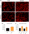
Immunostaining of RNA-binding protein and mRNA Processing Factor-positive (RBPMS+) cells for the analysis of retinal ganglion cell (RGC) density in flat-mount retinas. (a) RBPMS+ RGCs in central (A,B) and peripheral areas (C,D) of control (CTRL) and Epicolin formulation-treated mice. Quantification was carried out on the whole retina (b) and on both central and peripheral areas of the retina (c). Scale bar corresponds to 50 μm. The data were plotted as means ± SEMs (n = 12 retinas per group). * p ≤ 0.05 vs. CTRL; ° p ≤ 0.05 vs. CTRL c; † p ≤ 0.05 vs. CTRL p. Unpaired t-test. CTRL central (CTRL c), CTRL periphery (CTRL p), Epicolin central (Epicolin c), and Epicolin periphery (Epicolin p).
Figure 9
Figure 9
Epicolin formulation effect on neurotrophic and inflammatory biomarkers. (a) Brain-derived neurotrophic factor (BDNF), (b) interleukin-1 beta (IL-1β), and (c) tumor necrosis factor-alpha (TNF-α) expression of Epicolin-treated mice compared to the control (CTRL) group. Each bar represents the mean value ± SEM (n = 10 per group). * p ≤ 0.05 vs. CTRL. Unpaired t-tests were performed.

References

    1. Cheung W., Guo L., Cordeiro M.F. Neuroprotection in Glaucoma: Drug-Based Approaches. Optom. Vis. Sci. 2008;85:406–416. doi: 10.1097/OPX.0b013e31817841e5. - DOI - PMC - PubMed
    1. Bou Ghanem G.O., Wareham L.K., Calkins D.J. Addressing Neurodegeneration in Glaucoma: Mechanisms, Challenges, and Treatments. Prog. Retin. Eye Res. 2024;100:101261. doi: 10.1016/j.preteyeres.2024.101261. - DOI - PubMed
    1. Soto I., Howell G.R. The Complex Role of Neuroinflammation in Glaucoma. Cold Spring Harb. Perspect. Med. 2014;4:a017269. doi: 10.1101/cshperspect.a017269. - DOI - PMC - PubMed
    1. Adornetto A., Rombolà L., Morrone L.A., Nucci C., Corasaniti M.T., Bagetta G., Russo R. Natural Products: Evidence for Neuroprotection to Be Exploited in Glaucoma. Nutrients. 2020;12:3158. doi: 10.3390/nu12103158. - DOI - PMC - PubMed
    1. Davis B.M., Crawley L., Pahlitzsch M., Javaid F., Cordeiro M.F. Glaucoma: The Retina and Beyond. Acta Neuropathol. 2016;132:807–826. doi: 10.1007/s00401-016-1609-2. - DOI - PMC - PubMed

LinkOut - more resources