. 2024 Dec 25;17(1):11.
doi: 10.3390/v17010011.

Exploration of the Role of Cyclophilins in Established Hepatitis B and C Infections

Affiliations

Exploration of the Role of Cyclophilins in Established Hepatitis B and C Infections

Jennifer Molle et al. Viruses. .

Abstract

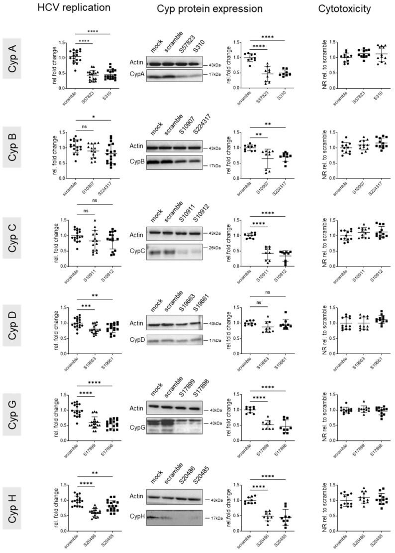
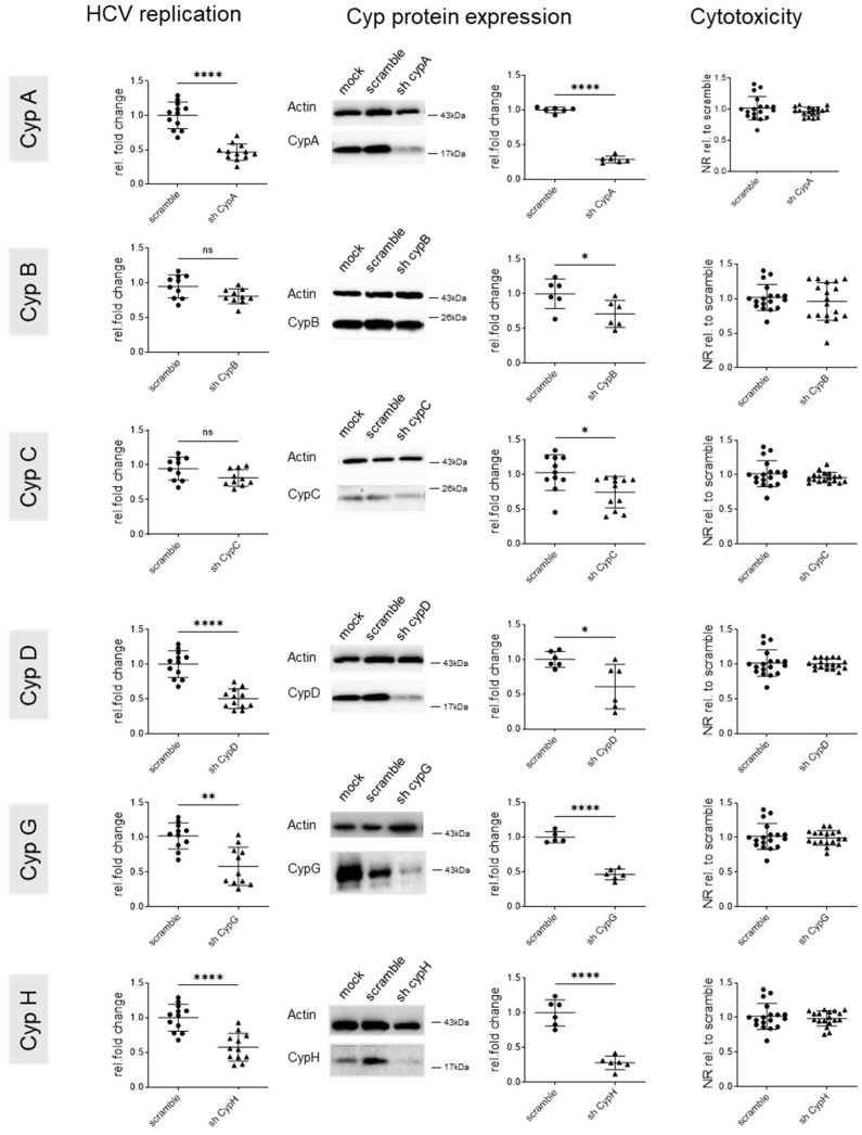
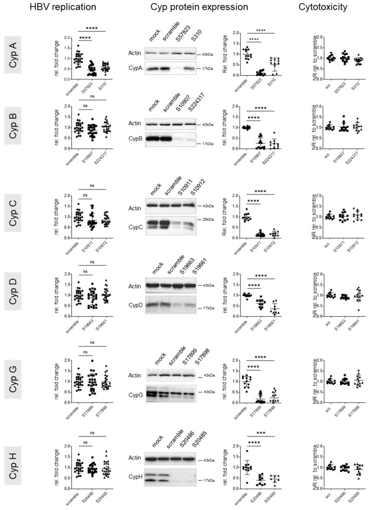
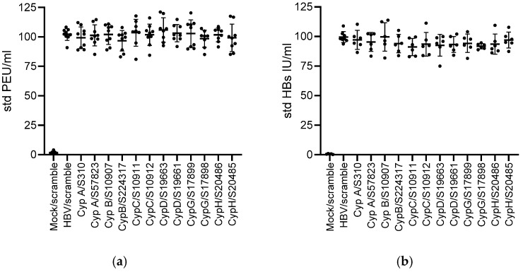
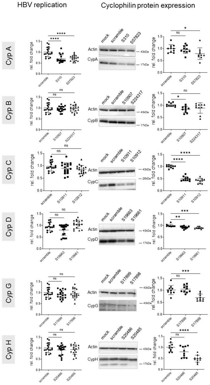
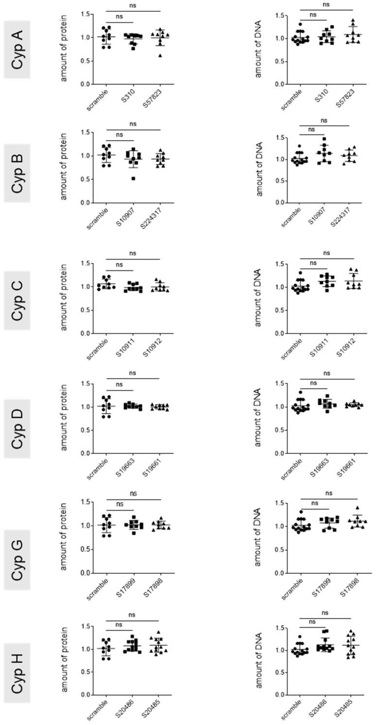
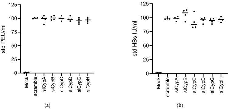
Cyclophilin (Cyp) inhibitors are of clinical interest in respect to their antiviral activities in the context of many viral infections including chronic hepatitis B and C. Cyps are a group of enzymes with peptidyl-prolyl isomerase activity (PPIase), known to be required for replication of diverse viruses including hepatitis B and C viruses (HBV and HCV). Amongst the Cyp family, the molecular mechanisms underlying the antiviral effects of CypA have been investigated in detail, but potential roles of other Cyps are less well studied in the context of viral hepatitis. Furthermore, most studies investigating the role of Cyps in viral hepatitis did not investigate the potential therapeutic effects of their inhibition in already-established infections but have rather been performed in the context of neo-infections. Here, we investigated the effects of genetically silencing Cyps on persistent HCV and HBV infections. We confirm antiviral effects of CypA and CypD knock down and demonstrate novel roles for CypG and CypH in HCV replication. We show, furthermore, that CypA silencing has a modest but reproducible impact on persistent HBV infections in cultured human hepatocytes.

Keywords: anti-viral treatment; cyclophilin; hepatitis virus; liver.


Conflict of interest statement

The authors declare no conflicts of interest. The funders had no role in the design of this study; in the collection, analyses, or interpretation of data; in the writing of this manuscript; or in the decision to publish the results.

Figures

Figure 1
Figure 1
HCV infection at 3 days post infection. Huh7.5 cells were infected with HCV at an MOI of 0.1 and fixed for immunofluorescence analysis 3 days later. Green: anti-HCV patient serum; blue: DAPI staining. A representative image is shown; scale bar: 50 µm.
Figure 2
Figure 2
Effect of genetic cyclophilin knock down (siRNA) on intracellular HCV replication. Huh7.5 cells were infected at an MOI of 0.1. Then, 72 h later, when HCV replication was well established, cells were transfected with siRNAs targeting the indicated Cyps and harvested 5 days after transfection. Left panels: intracellular HCV replication determined by RTqPCR with GUS as housekeeping gene and standardized to scramble controls. Middle panels: Cyp expression was assessed by Western blotting, quantified using ImageJ, standardized to actin, and data were expressed relative to the scramble conditions. Representative Western blots are shown. Right panels: cytotoxicity was assessed by neutral red staining with data standardized to the scramble control. Means +/− STD, n = 3, with each experiment performed with at least three culture wells and each well representing a datapoint. One-way anova. * p < 0.05, ** p < 0.01, and *** p < 0.001; **** p < 0.0001.
Figure 3
Figure 3
Effect of genetic cyclophilin knock down (shRNA) on intracellular HCV replication. Huh7.5 cells were infected at an MOI of 0.1. Then, 72 h later, when HCV replication was well established, cells were transfected with shRNA vectors targeting the indicated Cyps and harvested 5 days after transfection. Left panels: intracellular HCV replication was determined by RTqPCR using GUS as a housekeeping gene. Data were standardized to HCV RNA levels observed in scramble controls. Middel panels: Cyp expression was assessed by Western blotting, quantified using ImageJ, standardized to actin, and data were expressed relative to the scramble conditions. Representative Western blots are shown. Right panels: cytotoxicity was assessed by neutral red staining with data standardized to scramble control. Means +/− STD, n = 3, with each experiment performed with at least two culture wells and each well representing a datapoint. One-way anova. * p < 0.05, ** p < 0.01, and **** p < 0.0001.
Figure 4
Figure 4
Effect of genetic cyclophilin knock down on intracellular HBV replication. HepG2-hNTCP cells were seeded at 4.105/well into 12 wells; cells were infected at day 3 at an MOI of 100 and transfected at day 10 with the indicated siRNAs. Cells were harvested 5 days after transfection to assess HBV replication and Cyp expression. Left panel: intracellular HBV replication was determined by qPCR using RPLP0 as a housekeeping gene. Results are expressed relative to scramble; middle panel: Cyp expression was assessed by Western blotting, quantified using ImageJ, standardized to actin, and data were expressed relative to the scramble conditions. Representative Western blots are shown. Right panel: cytotoxicity was assessed by neutral red staining with data standardized to the scramble control. Means +/− STD, n = 3, with each experiment performed with a minimum of three culture wells and each well representing a datapoint. One-way anova. *** p < 0.001 and **** p < 0.0001.
Figure 5
Figure 5
HBs and HBe antigen secretion are not affected by Cyp knock down. HepG2-hNTCP cells were seeded at 4.105/well into 12 wells, DMSO was added 1 day later for 24 h. Cells were then infected at an MOI of 100 and transfected 7 days later with the indicated siRNAs. Cell supernatants were harvested at the time of transfection at day 7 and 5 days after transfection to assess (a) HBe (PEIU/mL) and (b) HBs (UI/mL) antigen secretion. Data were standardized to values obtained for the HBV/scramble control at day 7 (time of transfection). Means +/− STD, n = 5. One-way anova: no statistically significant difference was observed between the conditions.
Figure 6
Figure 6
Effect of genetic Cyp knock down on intracellular HBV replication in PHH. PHHs were infected (MOI 250) and transfected 7 days later with the siRNAs targeting the indicated Cyps. Cells were harvested 5 days after transfection. Left panels: intracellular HBV replication quantified by qPCR with RPLP0 as a housekeeping gene. Right panels: Cyclophilin expression by Western blotting, quantified with ImageJ, standardized to actin, and data were expressed relative to the scramble. Representative Western blots are shown. Means +/− STD, n = 3, with each experiment performed with at least three culture wells with each well representing a datapoint. One-way anova. * p < 0.05, ** p < 0.01, *** p < 0.001 and **** p < 0.0001.
Figure 7
Figure 7
Cytotoxicity of genetic cyclophilin knock down by siRNA. PHHs were seeded at 1.106/well into 12 wells. DMSO was added 1 day later for 24 h. Cells were then infected at an MOI of 250 and transfected 7 days later with the indicated siRNAs. Cells were harvested 5 days after transfection. Total protein (left panel) and DNA (right panel) were quantified as the read out for cell toxicity. n = 3, means +/− STD, with each experiment performed with at least three culture wells with each well representing a datapoint; One-way anova.
Figure 8
Figure 8
HBs and HBe antigen secretion are not affected by Cyp knock out. PHHs were seeded at 1.106/well into 12 wells. DMSO was added 1 day later for 24 h. Cells were then infected at an MOI of 250 and transfected 7 days later with the mixtures of siRNAs (as used in Figure 6), targeting the indicated siRNAs. Cell supernatants were harvested at the time of transfection at day 7 and 5 days after transfection to assess (a) HBe (PEIU/mL) and (b) HBs (UI/mL) antigen secretion. Data were standardized to values obtained for the HBV/scramble control at day 7 (time of transfection). Means +/− STD, n = 4. One-way anova: no statistically significant difference was observed between the conditions.

Similar articles

References

    1. Stamnes M.A., Rutherford S.L., Zuker C.S. Cyclophilins: A new family of proteins involved in intracellular folding. Trends Cell Biol. 1992;2:272–276. doi: 10.1016/0962-8924(92)90200-7. - DOI - PubMed
    1. Nigro P., Pompilio G., Capogrossi M.C. Cyclophilin a: A key player for human disease. Cell Death Dis. 2013;4:e888. doi: 10.1038/cddis.2013.410. - DOI - PMC - PubMed
    1. Kim J., Choi T.G., Ding Y., Kim Y., Ha K.S., Lee K.H., Kang I., Ha J., Kaufman R.J., Lee J., et al. Overexpressed cyclophilin b suppresses apoptosis associated with ros and Ca2+ homeostasis after er stress. J. Cell Sci. 2008;121:3636–3648. doi: 10.1242/jcs.028654. - DOI - PMC - PubMed
    1. Elrod J.W., Molkentin J.D. Physiologic functions of cyclophilin d and the mitochondrial permeability transition pore. Circ. J. 2013;77:1111–1122. doi: 10.1253/circj.CJ-13-0321. - DOI - PMC - PubMed
    1. Rajiv C., Davis T.L. Structural and functional insights into human nuclear cyclophilins. Biomolecules. 2018;8:161. doi: 10.3390/biom8040161. - DOI - PMC - PubMed

Publication types

LinkOut - more resources