. 2025 Mar;301(3):108221.
doi: 10.1016/j.jbc.2025.108221. Epub 2025 Jan 23.

Downregulation of the phosphatase PHLPP1 contributes to NNK-induced malignant transformation of human bronchial epithelial cells (HBECs)

Affiliations

Downregulation of the phosphatase PHLPP1 contributes to NNK-induced malignant transformation of human bronchial epithelial cells (HBECs)

Xuelei Liu et al. J Biol Chem. 2025 Mar.

Abstract

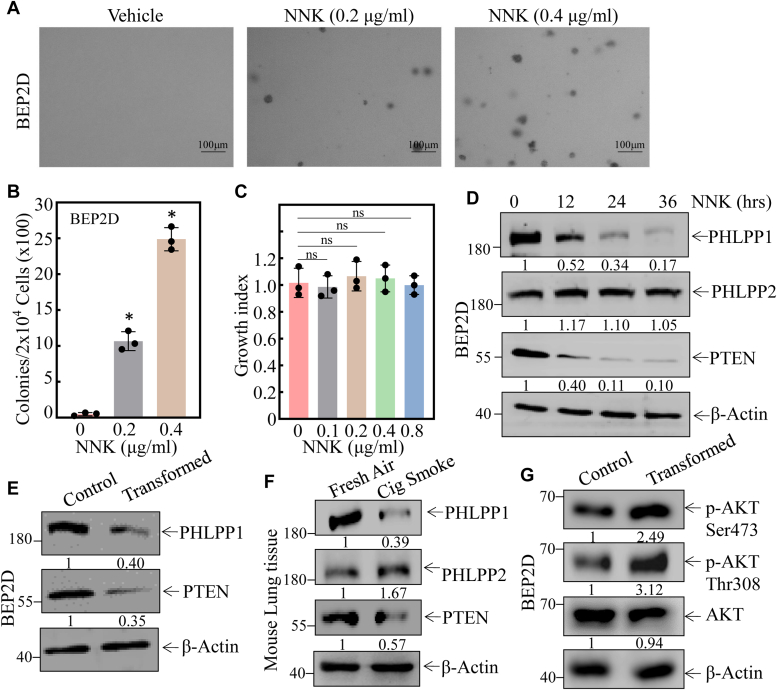
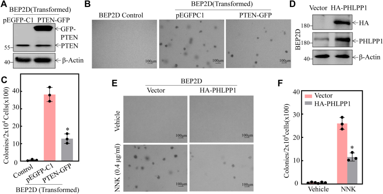
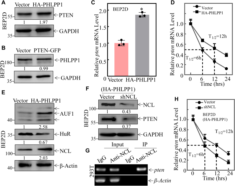
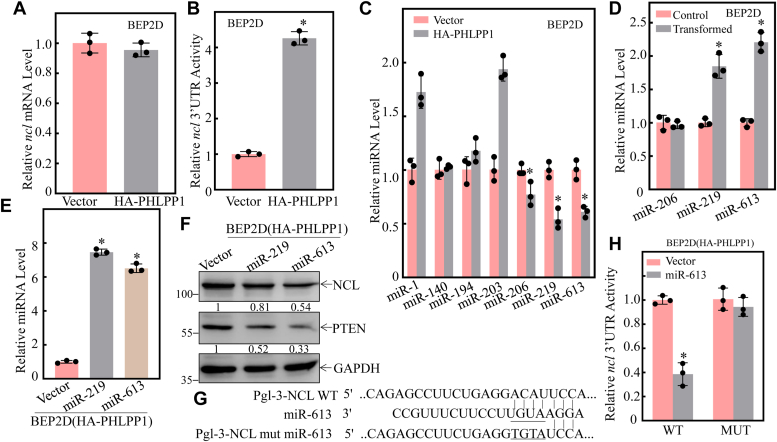
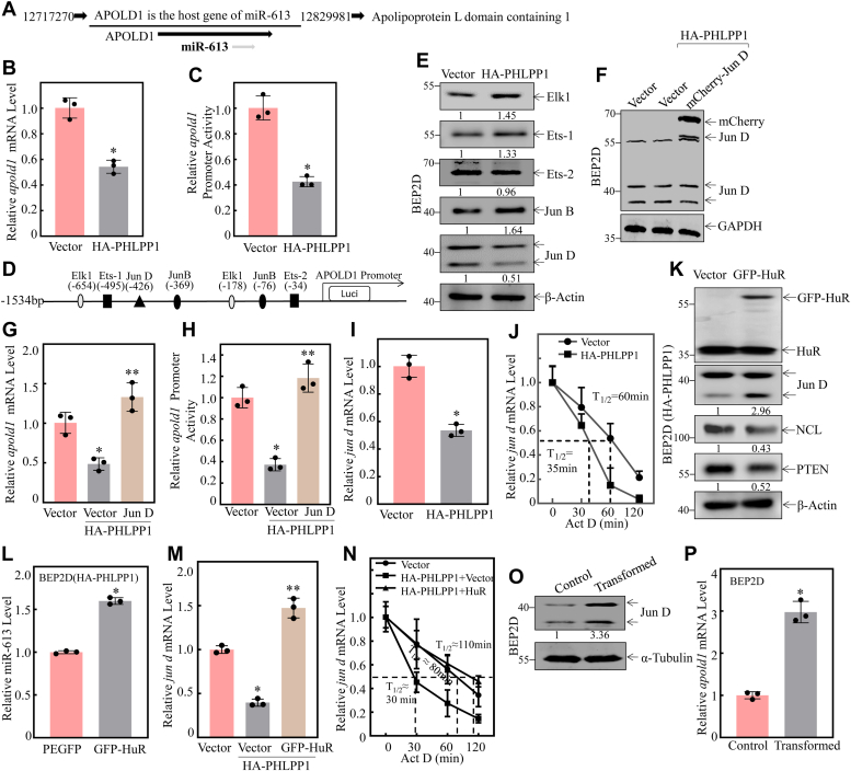
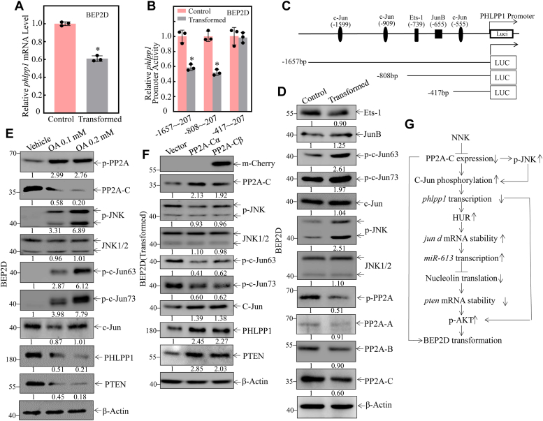
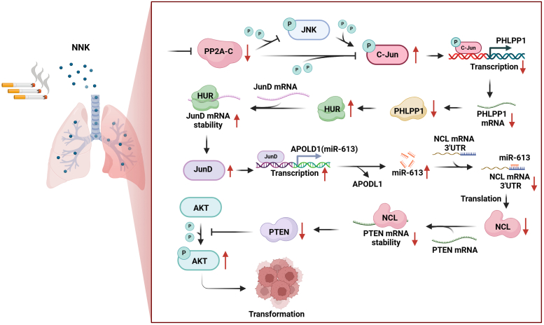
Cigarette smoking (CS) is one of the greatest health concerns, which can cause lung cancer. 4-(Methylnitrosamino)-1-(3-pyridyl)-1-butanone (NNK), a tobacco-specific nitrosamine, has been well-documented for its carcinogenic activity in both epidemiological and laboratory studies. PH domain leucine-rich repeat protein phosphatase 1 (PHLPP1) and phosphatase and tensin homolog (PTEN) are two well-known phosphatase tumor suppressors that have been reported to be downregulated in human lung cancer tissues. However, the effect of NNK exposure on the expression of PHLPP1 and PTEN is unknown, and such effects may be early events leading to lung carcinogenesis. We explored this question in current studies and found that exposure of human bronchial epithelial BEP2D cells to NNK resulted in cell malignant transformation accompanied by a remarkable downregulation of PHLPP1 and PTEN. Such downregulation of PHLPP1 and PTEN was also consistently observed in vivo in Cigarette Smoking-exposed mouse lung tissues. Our studies further showed that overexpression of PHLPP1 or PTEN alleviated NNK-induced BEP2D cell transformation. Ectopic expression of PHLPP1 promoted PTEN protein expression, while PTEN overexpression did not affect PHLPP1 expression. Mechanistic studies showed that NNK was able to inhibit PP2A-C activity, which directly attenuated c-Jun phosphorylation at Ser63/73, and subsequently inhibited the PHLPP1 transcription and expression. Further, the downregulation of PHLPP1 led to a reduction of pten mRNA stability and expression through the HUR/Jun D/miR-613/NCL axis. Taken together, our studies advance the field of tobacco-induced lung cancer research by uncovering new mechanistic insights and identifying a novel molecular axis that could inform future therapeutic strategies. It also adds a new dimension by exploring the interaction between PHLPP1 and PTEN in the context of tobacco carcinogen exposure.

Keywords: NNK; PHLPP1; PP2AC; PTEN; lung carcinogenesis; miR-613.


Conflict of interest statement

Conflict of interest The authors declare that they have no conflicts of interest with the contents of this article.

Figures

Figure 1
Figure 1
Downregulation of PTEN and PHLPP1 expression was observed following NNK exposure in vivo and in vitro.A and B, chronic exposure to NNK caused BEP2D (Nonsense) cells to acquire anchorage-independent growth in soft agar, a hallmark of malignant cellular transformation. The number of colonies was counted and presented as colonies per 104 seeded cells. The bars shown are mean ± SD from three independent experiments. ∗Significant difference relative to vehicle control (p < 0.05). C, the cell growth index of BEP2D cells was evaluated after NNK exposure through the CCK-8 assay. ns p > 0.05. D, Western Blot was employed to determine the expression of phosphatases, including PHLPP1, PHLPP2, and PTEN. β-actin was measured as an internal control. E, the relative levels of PHLPP1, PHLPP2, and PTEN were determined in BEP2D(Control) and BEP2D(transformed) cells. β-actin was measured as an internal control. F, Western Blot assay was performed to evaluate the expression of PHLPP1, PHLPP2, and PTEN in fresh air-exposed mouse lung tissues and cigarette smoking-exposed mouse lung tissues (FA, fresh air; CS, cigarette smoking; n = 5). β-actin was measured as an internal control. G, Western Blot was used to detect the expression of AKT phosphorylation of Ser473 and Thr308 sites in NNK-induced transformed BEP2D cells. β-actin was measured as an internal control. All experiments were performed at least three replicates.
Figure 2
Figure 2
PTEN and PHLPP1 played a vital role in the NNK-induced malignant transformation of BEP2D cells.A, overexpress construct of PTEN was stably transfected into BEP2D(transformed) cells. Western Blot was used to detect the knockdown efficiency of PTEN protein. B and C, the indicated cells were subjected to soft agar assay. (B) The number of colonies was scored and presented as colonies per 104 seeded cells (C). The asterisk (∗) indicates a significant decrease in comparison to BEP2D(PEGFPC1) cells (p < 0.05). D, overexpress construct of the PHLPP1 was stably transfected into BEP2D cells. Western Blot was used to determine HA-PHLPP1 protein expression level. E and F, the BEP2D(Vector) and BEP2D(HA-PHLPP1) cells were exposed to 400 μg/ml NNK and then subjected to soft agar assay. E, the number of colonies was scored and presented as colonies per 104 seeded cells. F, the asterisk (∗) indicates a significant change (p < 0.05). All experiments were performed at least three replicates.
Figure 3
Figure 3
PHLPP1 upregulates pten mRNA stability through upregulating NCL in BEP2D(HA-PHLPP1) cells compared to BEP2D(Vector) cells.A, cell extracts obtained from the BEP2D(Vector) and BEP2D(HA-PHLPP1) transfectants were subjected to Western blot for determination of the PTEN protein expression. β-Actin was used as protein loading control. B, cell extracts obtained from the BEP2D(Vector) and BEP2D(PTEN-EGFPC1) transfectants were subjected to Western blot to determination of the PHLPP1 protein expression. β-Actin was used as the protein loading control. C, relative pten mRNA levels in cells were evaluated using real-time PCR in BEP2D(Vector) and BEP2D(PTEN-EGFPC1) stably transfectants. Bars represent means ± SD from three independent experiments. The asterisk (∗) indicates a significant change (p < 0.05). β-actin was used as the mRNA loading control. D, relative pten mRNA stability was evaluated by real-time PCR in BEP2D(Vector) vs. BEP2D(HA-PHLPP1) cells after treatment with Act D for indicated times. β-actin was used as the mRNA loading control. E, the indicated cell extracts were subjected to Western Blot to determination of AUF1, NCL, and HUR protein expression. β-Actin was used as the protein loading control. F, sh-NCL or its control vector was transfected into BEP2D(HA-PHLPP1) cells, and the indicated stable transfectants were subjected to Western blot to determination of NCL and PTEN protein expression as indicated. GAPDH was used as protein loading control. G, RNA immunoprecipitation assay using an anti-NCL antibody to test the interaction of NCL with pten mRNA in 293T cells. H, relative pten mRNA stability was evaluated by real-time PCR in the indicated cell cells after treatment with Act D for indicated times. β-actin was used as the mRNA loading control. All experiments were performed at least three replicates.
Figure 4
Figure 4
miR-613 binds to the 3′-UTR of ncl mRNA and downregulates its protein expression in the NNK-induced transformaed BEP2D cells.A, relative NCL mRNA level was evaluated by real-time PCR in BEP2D(Vector) vs. BEP2D(HA-PHLPP1) cells. B, pMIR-NCL 3′-UTR reporters were transiently transfected into the BEP2D(Vector) or BEP2D(HA-PHLPP1) cells, and luciferase activity was evaluated. The asterisk (∗) indicates a significant change (p < 0.05). C, qPCR was performed to determine the expression of miRNAs in BEP2D (Vector) vs. BEP2D(HA-PHLPP1) cells. The asterisk (∗) indicates a significant change (p < 0.05). The U6 was used as the loading control. D, qPCR was performed to determine the effect of miRNA expression in BEP2D(Control) vs. BEP2D(transformed) cells. The U6 was used as the loading control. E, miR-219 and miR-613 overexpression was identified in BEP2D cells. The asterisk (∗) indicates a significant increase in BEP2D(miR-219) cells vs. BEP2D(Vector) or BEP2D(miR-613) cells vs. BEP2D(Vector) cells (p < 0.05). The U6 was used as the loading control. F, NCL and PTEN expressions were analyzed by Western blot in BEP2D(Vector) vs. BEP2D(miR-219) and BEP2D (miR-613) cells. GAPDH was used as the protein loading control. G, schematic of the miR-613 binding site in ncl mRNA 3′-UTR regions, and its mutants aligned with miR-613. H, the indicated cells were co-transfected with wild-type, mutant NCL 3′-UTR luciferase reporters, and pRL-TK, respectively. Luciferase activity of each transfectant was evaluated and results were presented as relative ncl 3′-UTR activity. All experiments were performed at least three replicates.
Figure 5
Figure 5
Increase in Jun D transcriptionally regulates miR-613 expression during NNK-induced malignant transformed BEP2D cells.A, APOLD1 is the host gene of miR-613. B, relative apold1 mRNA level was evaluated by real-time PCR in BEP2D(Vector) vs. BEP2D(HA-PHLPP1) cells. The β-actin was used as the mRNA loading control. C, the apold1 promoter-driven luciferase reporter constructs, together with TK, were stably transfected into BEP2D(Vector) vs. BEP2D(HA-PHLPP1), respectively. The transfected cells were extracted to evaluate the luciferase activity after 48 h, and the results are presented as relative apold1 promoter activity. The asterisk (∗) indicates a significant decrease of apold1 promoter transcriptional activity in BEP2D(HA-PHLPP1) compared to BEP2D(Vector) cells (p < 0.05). D, potential transcriptional factor binding sites in the APOLD1 promoter region (−1534∼+0) were analyzed using the TRANSFAC 8.3 engine online. E, Western blotting was used to detect Elk1, Ets-1, Ets-2, JunB, and JunD protein expression in the cells. β-Actin was used as a protein loading control. F, Western blotting was used to detect the overexpression efficiency of Jun D protein expression in BEP2D(HA-PHLPP1) cells. GAPDH was used as a protein loading control. G, relative apod1 mRNA level was evaluated by real-time PCR in the indicated cells after Jun D overexpression. The asterisk (∗) indicates a significant decrease compared to BEP2D(Vector) cells (p < 0.05). The asterisk (∗∗) indicates a significant increase compared to BEP2D(HA-PHLPP1/Vector) cells (p < 0.001). The β-actin was used as the mRNA loading control. H, the apold1 promoter-driven luciferase reporter constructs, together with TK, were stably transfected into the indicated cells. The transfected cells were extracted to evaluate the luciferase activity after 48 h, and the results are presented as relative phlpp1 promoter activity. The asterisk (∗) indicates a significant decrease compared to BEP2D(Vector) cells (p < 0.05). The asterisk (∗∗) indicates a significant increase compared to BEP2D(HA-PHLPP1/Vector) cells (p < 0.001). I, relative jun d mRNA level was evaluated by real-time PCR in BEP2D(Control) vs. BEP2D(HA-PHLPP1) cells. The β-actin was used as the mRNA loading control. J, relative jun d mRNA stability was evaluated by real-time PCR in the indicated cell cells after treatment with Act D for indicated times. K, GFP-HUR or its control vector was transfected into BEP2D(HA-PHLPP1) cells, and the indicated stable transfectants were subjected to Western Blot to determine JunD, NCL, and PTEN protein expression as indicated. β-Actin was used as the protein loading control. L, qPCR was performed to assess the effect of overexpression GFP-HUR on miR-613 expression in BEP2D(HA-PHLPP1) cells. The asterisk (∗) indicates a significant change (p < 0.05). The U6 was used as the miRNA loading control. M, relative jun d mRNA stability was evaluated by real-time PCR in the indicated cell cells. The asterisk (∗) indicates a significant inhibition compared to DEP2D(Vector) cells (p < 0.05); The asterisk (∗∗) indicates a significant increase compared to BEP2D(HA-PHLPP1/Vector) cells (p < 0.001). The β-actin was used as the mRNA loading control. N, relative jun d mRNA stability was evaluated by real-time PCR in the indicated cell cells after treatment with Act D for indicated times. O, Western blotting was used to detect Jun D protein expression in BEP2D transformed cells. α-Tubulin was used as a protein loading control. P, relative apold1 mRNA level was evaluated by real-time PCR in BEP2D transformed cells. The β-actin was used as the mRNA loading control. All experiments were performed at least three replicates.
Figure 6
Figure 6
NNK increased phosphorylation of c-Jun at Ser-63/Ser-73 through down-regulating PP2A-C and up-regulating phosphorylation of JNK.A, relative phlpp1 mRNA level was evaluated by real-time PCR in BEP2D(Control) vs. BEP2D(transformed) cells. β-actin was used as the mRNA loading control. B, the various phlpp1 promoter-driven luciferase reporter constructs, together with TK, were stably transfected into BEP2D (Control) and BEP2D (transformed) cells, respectively. The transfected cells were extracted to evaluate the luciferase activity after 48 h, and the results are presented as relative phlpp1 promoter activity. The asterisk (∗) indicates a significant decrease in comparison to untransformed control cells (p < 0.05). C, a schematic illustration of the construction of phlpp1 promoter-driven luciferase reporter constructs. D, Western blotting was used to detect Ets-1, JunB, phosphorylated c-Jun at Ser63, phosphorylated c-Jun at Ser73, phosphorylated JNK, JNK, phosphorylated PP2A, PP2A-A, PP2A-B, PP2A-C subunit and c-Jun protein expression in the indicated cells. β-Actin was used as a protein loading control. E, BEP2D cells (2 × 105) were seeded into each well of 6-well plates. After synchronization, cells were treated with the indicated concentration of okadaic acid (OA) for 8 h in a traditional medium and then extracted for Western blotting to determine the protein levels of phosphorylated PP2A, PP2A-C subunit phosphorylated JNK, JNK1/2, phosphorylated c-Jun at Ser63, phosphorylated c-Jun at Ser73 and c-jun. β-Actin was used as a protein loading control. F, overexpression of PP2A-Cα and PP2A-Cβ in BEP2D(transformed) cells and Western blotting were used to detect phosphorylated PP2A, PP2A-C subunit phosphorylated JNK, JNK1/2, phosphorylated c-Jun at Ser63, phosphorylated c-Jun at Ser73 and c-jun protein expression in the cells. β-Actin was used as the protein loading control. G, the proposed mechanisms underlying the downregulation of PHLPP1 contribute to the NNK-induced malignant transformation of human bronchial epithelial cells through regulating pten mRNA stability. All experiments were performed at least three replicates.
Figure 7
Figure 7
The schematic diagram of NNK-induced malignant transformation of human bronchial epithelial cells. Briefly, NNK inhibited PP2A-C activity, which directly attenuated c-Jun phosphorylation at Ser63/73, and subsequently inhibited the PHLPP1 transcription and expression. The downregulation of PHLPP1 reduced pten mRNA stability and expression through the HUR/Jun D/miR-613/NCL axis, resulting in malignant transformation of human bronchial epithelial cells.

Similar articles

References

    1. Schuller H.M. Mechanisms of smoking-related lung and pancreatic adenocarcinoma development. Nat. Rev. Cancer. 2002;2:455–463. - PubMed
    1. Chen X., Zhang W., Liu R., Zhu Z., Gong M., Wang Q., et al. NNK from tobacco smoking enhances pancreatic cancer cell stemness and chemoresistance by creating a β2AR-Akt feedback loop that activates autophagy. Mol. Oncol. 2022;16:2881–2895. - PMC - PubMed
    1. Shen J., Xu L., Owonikoko T.K., Sun S.Y., Khuri F.R., Curran W.J., et al. NNK promotes migration and invasion of lung cancer cells through activation of c-Src/PKCι/FAK loop. Cancer Lett. 2012;318:106–113. - PMC - PubMed
    1. Alvarez-Garcia V., Tawil Y., Wise H.M., Leslie N.R. Mechanisms of PTEN loss in cancer: it's all about diversity. Semin. Cancer Biol. 2019;59:66–79. - PubMed
    1. Lemoine K.A., Fassas J.M., Ohannesian S.H., Purcell N.H. On the PHLPPside: emerging roles of PHLPP phosphatases in the heart. Cell Signal. 2021;86 - PMC - PubMed

MeSH terms